 Ghatkopar (East), Mumbai)


Home > Interventional radiology > Liver Tumors
Liver Tumor Treatment in Mumbai
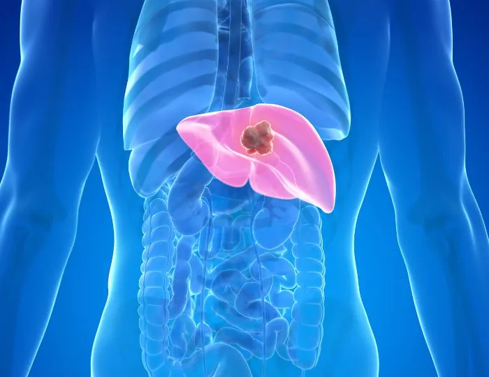
The liver is the most common site of both primary and secondary (metastatic) malignancy in the abdomen. Whether arising from hepatocytes (hepatocellular carcinoma), bile ducts (cholangiocarcinoma), or spread from colorectal, breast, lung, or neuroendocrine primaries, liver Tumor demand early, precise, and multidisciplinary intervention.
At Shree Hospitals, Ghatkopar East, Mumbai, our interventional radiology team offers the full spectrum of minimally invasive liver-directed therapies , from transarterial chemoembolisation (TACE) and selective internal radiation therapy (SIRT/TARE with Y-90) to radiofrequency ablation (RFA), microwave ablation (MWA), and irreversible electroporation (IRE/NanoKnife) , all performed in a dedicated angio suite by specialists trained in hepatic oncology.
(24×7 Emergency Care)
(24×7 Emergency Care)
Our Approach
(24×7 Emergency Care)
Our approach in managing Liver Tumors
Step 1 — Multidisciplinary Tumour Board Review and BCLC Staging
Every liver tumour case at Shree Hospitals is presented at our multidisciplinary tumour board involving hepatology, medical oncology, surgical oncology, and interventional radiology before any treatment is initiated. Staging follows the Barcelona Clinic Liver Cancer (BCLC) system , the only staging system that integrates tumour burden, liver function (Child-Pugh score), and performance status into treatment allocation. BCLC Stage 0–A (very early and early): surgical resection or ablation with curative intent. BCLC Stage B (intermediate): TACE or TARE. BCLC Stage C (advanced, portal invasion or metastases): systemic therapy ± TARE. BCLC Stage D (end-stage): best supportive care. Liver function is assessed with Child-Pugh score (A/B/C) and MELD score; portal hypertension assessment via hepatic venous pressure gradient (HVPG) guides procedural risk stratification.
Step 2 — Advanced Imaging for Lesion Characterisation and Planning
All liver Tumor undergo triphasic CT or gadoxetate-enhanced MRI (Primovist/Eovist) for lesion characterisation. HCC demonstrates arterial enhancement with portal venous or delayed washout on dynamic imaging , this "washout" pattern is diagnostic and eliminates the need for biopsy in cirrhotic patients with lesions >1cm (LI-RADS 5 = HCC until proven otherwise). Cholangiocarcinoma shows progressive peripheral enhancement. Metastases show varied patterns depending on the primary. For TARE planning, 99mTc-MAA (macroaggregated albumin) SPECT-CT is performed to calculate the hepatopulmonary shunt fraction and plan Y-90 dosimetry , critical to prevent radiation pneumonitis.
Step 3 — Liver-Directed Therapy Delivery in Dedicated Angio Suite
Liver-directed procedures at Shree Hospitals are performed in a biplane digital subtraction angiography (DSA) suite under conscious sedation. Super-selective catheterisation of hepatic arterial branches supplying the tumour ,using 2.7Fr or 2.0Fr microcatheters — allows targeted delivery of embolic agents, chemotherapy, or Y-90 microspheres directly to the tumour while sparing surrounding parenchyma. For ablation (RFA, MWA, IRE), real-time CT or ultrasound guidance is used for probe placement with 1cm ablation margin.
Step 4 — Portal Vein Embolisation for Surgical Preparation
When surgical resection is planned but the future liver remnant (FLR) volume is inadequate (<25–30% in normal liver, <40% in cirrhotic liver), portal vein embolisation (PVE) is performed to induce hypertrophy of the FLR before surgery. PVE involves percutaneous transhepatic embolisation of portal vein branches supplying the segments to be resected, diverting portal flow to the segments that will remain. CT volumetry at 4–6 weeks post-PVE confirms FLR hypertrophy; resection is then performed when the FLR is adequate.
Step 5 — Response Assessment and Ongoing Surveillance
Treatment response is assessed by mRECIST criteria (modified RECIST) at 4–6 weeks post-TACE/TARE and 3 months post-ablation , measuring the viable, enhancing tumour component rather than total tumour size. Complete response (CR): no arterial enhancement in the treated area. Partial response (PR): ≥30% reduction in enhancing diameter. Progressive disease: ≥20% increase or new lesion. Patients with complete ablative response enter surveillance with triphasic CT or MRI every 3 months. Repeat TACE or bridging to transplant is planned based on response and evolving tumour burden.
How to identify Liver Tumors?
Liver Tumor are particularly insidious because the liver has no pain fibres within its parenchyma ,pain only occurs when the tumour stretches the liver capsule or invades adjacent structures. By the time symptoms appear, the tumour is often large or multifocal.
Common symptoms that may indicate liver Tumor include persistent right upper quadrant or epigastric discomfort or a feeling of fullness after small meals, unexplained weight loss and loss of appetite over weeks to months, progressive jaundice (yellow discolouration of skin and eyes) indicating biliary compression or liver function deterioration, abdominal swelling from ascites (fluid accumulation) in the setting of underlying cirrhosis, fatigue and weakness disproportionate to other illness, and in advanced cases, a palpable mass in the right upper abdomen.
In patients with known cirrhosis from hepatitis B, hepatitis C, alcohol-related liver disease, or NAFLD, HCC surveillance with 6-monthly ultrasound and AFP testing is standard protocol , the goal is to detect Tumor at BCLC Stage 0 or A when curative treatment is possible.
Urgency indicators requiring immediate evaluation: sudden worsening of ascites (may indicate tumour rupture or portal vein thrombosis), haematemesis (variceal bleed from portal hypertension exacerbated by tumour), rapid deterioration in liver function in a previously stable cirrhotic patient, and new onset confusion or encephalopathy in a previously stable patient.
Laboratory markers: AFP (alpha-fetoprotein) >400 ng/mL is strongly suggestive of HCC but not present in all cases (elevation in only 60–70% of HCC). AFP-L3 fraction and des-gamma-carboxyprothrombin (DCP/PIVKA-II) add sensitivity. For cholangiocarcinoma: CA 19-9 and CEA. For colorectal metastases: CEA and CA 19-9.
Important FAQs: Liver Tumors
What is the difference between primary liver cancer and liver metastases?
Primary liver cancer — hepatocellular carcinoma (HCC) or cholangiocarcinoma , originates in liver cells or bile duct cells. Liver metastases are cancers that started elsewhere (colorectal, breast, lung, neuroendocrine) and spread to the liver through the bloodstream. The treatment approach differs significantly: HCC in cirrhotic patients requires liver function–adjusted staging (BCLC) and liver-directed therapies; metastases treatment is guided by the primary cancer's biology and extent of spread. Both types are treatable at Shree Hospitals with minimally invasive image-guided techniques.
Is TACE a surgery? Will I need general anaesthesia?
TACE is not surgery. It is performed in an interventional radiology suite through a small puncture in the groin (femoral artery) or wrist (radial artery), under conscious sedation and local anaesthesia , similar to a coronary angiogram. Most patients are awake and comfortable during the procedure, which takes 60–90 minutes. Post-embolisation syndrome (fever, nausea, abdominal pain) is expected for 48–72 hours and is managed with supportive care.
What is post-embolisation syndrome and is it dangerous?
Post-embolisation syndrome (PES) is the expected inflammatory response to ischaemic tumour necrosis following TACE. It manifests as low-grade fever (37.5–38.5°C), right upper quadrant pain, nausea, and malaise, typically lasting 3–7 days. PES is managed with paracetamol, anti-emetics, and adequate hydration , it is not an infection and does not require antibiotics. Severe fever (>38.5°C persisting beyond 7 days), jaundice, or signs of liver failure warrant reassessment and are differentiated from expected PES by clinical and biochemical monitoring.
How many TACE sessions will I need?
TACE is typically performed in a series of sessions, with MRI assessment 4–6 weeks after each session. The number of sessions depends on treatment response, tumour characteristics, and liver function. Patients with complete response after 1–2 sessions enter surveillance. Those with partial response may receive repeat TACE or be switched to TARE or systemic therapy. Repeat TACE is avoided if liver function deteriorates (Child-Pugh B progression) or if the tumour demonstrates TACE refractoriness (no objective response after 2 sessions).
Can liver Tumor be completely cured with ablation?
For small (<3cm) HCC or solitary liver metastases in surgically unfit patients, RFA and MWA achieve complete ablation rates of 85–95%, with 5-year survival comparable to surgical resection in selected patients. "Cure" requires sustained complete response on follow-up imaging with no local recurrence. Local tumour progression after ablation occurs in 3–12% of cases for lesions <3cm and is treated with repeat ablation or alternative therapy. Ablation is considered curative in intent for BCLC Stage 0–A HCC when surgical resection is not feasible.
Treatments for Liver Tumors at Shree Hospitals
TACE (Transarterial Chemoembolisation)
Conventional TACE (cTACE) uses Lipiodol as a drug carrier combined with doxorubicin or cisplatin, followed by gelfoam or PVA particle embolisation. Drug-eluting bead TACE (DEB-TACE) loads calibrated microspheres (DC Beads, HepaSphere) with doxorubicin for controlled drug release. TACE is the standard of care for BCLC Stage B (intermediate) HCC and is used as a bridge to transplant for early-stage Tumor exceeding Milan criteria. It is performed as a day-care procedure with overnight monitoring.
TARE / SIRT (Y-90 Selective Internal Radiation Therapy)
Y-90 TARE (transarterial radioembolisation) uses resin (SIR-Spheres) or glass (TheraSphere) microspheres loaded with yttrium-90, a pure beta-emitter with 2.67-day half-life and 2.5mm tissue penetration. Selective delivery via hepatic artery delivers an ablative radiation dose (120–150 Gy to tumour) with minimal systemic toxicity. TARE is preferred over TACE in patients with portal vein thrombosis (tumour thrombus), large or bilobar disease, and for downstaging HCC into transplant criteria. Available at Shree Hospitals as part of our advanced oncology programme.
Thermal Ablation (RFA & MWA)
Radiofrequency ablation (RFA) uses alternating current at 400–500 kHz to generate frictional heat (90–100°C), causing coagulative necrosis in a 3–5cm zone. Microwave ablation (MWA) uses 2.45 GHz microwave energy for faster heating, larger ablation zones (5–6cm), and superior performance in perivascular Tumor where the heat sink effect limits RFA. Both are performed under ultrasound or CT guidance and achieve complete ablation in Tumor <3cm with 5-year local recurrence rates comparable to surgical resection. Ideal for BCLC Stage 0–A patients unfit for surgery.
IRE / NanoKnife (Irreversible Electroporation)
NanoKnife IRE is the only non-thermal ablation modality, using high-voltage microsecond electrical pulses to create permanent nanopores in cell membranes — causing cell death without heat generation. This allows safe ablation of Tumor adjacent to bile ducts, portal vein, hepatic artery, and other critical structures where thermal ablation would cause collateral damage. IRE is used for centrally located HCC, perihilar cholangiocarcinoma, and liver metastases abutting major vessels. At Shree Hospitals, IRE is performed under general anaesthesia with cardiac synchronisation to prevent arrhythmia.
Liver Tumors Specialists in Mumbai
Now at Shree Hospitals

10+ years of experience
Dr. Swati Narurkar
Interventional Radiology

For Appointment/Query
1800-268-4000

Tele consultation/2nd Opinion
Schedule Consultation

News & Updates
Shree Hospitals Expands Paediatric Surgical Care With Latest Technology
Shree Hospitals Installs a Robotic-Assisted Surgical System for Better Patient Care


