 Ghatkopar (East), Mumbai)

Home > Interventional Radiology > Percutaneous Drainage (Abscess)
Percutaneous Abscess and Fluid Drainage In Mumbai
Image-Guided Drainage of Abscesses and Fluid Collections Without Open Surgery
Percutaneous drainage is a minimally invasive interventional radiology procedure that uses ultrasound or CT image guidance to drain abscesses, fluid collections, cysts, haematomas, and bilomas from within the body through a small skin puncture, without the need for open surgery. At Shree Hospitals, our specialist interventional radiologists perform percutaneous drainage as a safe, rapid, and effective treatment for abdominal, pelvic, liver, lung, and post-operative fluid collections, resolving infection and relieving pressure while avoiding the risks of general anaesthesia and major surgery.
Abscess or Fluid Collection Diagnosed? Ask About Percutaneous Drainage Today
Quick facts
Procedure Type : Image-Guided Needle Drainage of Abscess or Fluid Collection
Anaesthesia : Local Anaesthesia (most cases); Conscious Sedation if needed
Duration : 30 to 60 minutes
Hospital Stay : As per underlying condition (drain left in place 2 to 7 days)
Recovery Time : Rapid symptom relief; drain removed when drainage minimal
Support Team : Interventional radiologists, surgeons, infectious disease specialists
Key Benefit : Drains infection without open surgery avoids general anaesthesia
What is Percutaneous Abscess and Fluid Collection Drainage?
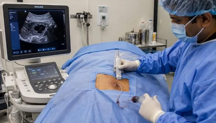
Percutaneous drainage is the image-guided insertion of a drainage catheter through the skin into an abscess, infected fluid collection, cyst, or haematoma to drain it externally. Using ultrasound or CT guidance, the interventional radiologist identifies the precise location and depth of the collection, plans the safest access route avoiding major blood vessels and organs, and then inserts a thin drainage catheter through a small skin incision directly into the collection. The infected fluid or pus drains through the catheter into an external bag. The catheter is secured in place and left until drainage is complete.
Percutaneous drainage is used for a wide variety of collections including abdominal and pelvic abscesses (post-operative, diverticular, appendiceal, Crohn's-related, gynaecological), liver abscesses (pyogenic and amoebic), splenic abscesses, retroperitoneal collections, pleural empyemas (infected fluid around the lung), lung abscesses, pancreatic pseudocysts and walled-off necrosis after pancreatitis, pericardial effusions, and post-traumatic haematomas. It can also be used for diagnostic aspiration - taking a small fluid sample for microbiological culture to identify the causative organism and guide antibiotic therapy.
At Shree Hospitals, percutaneous drainage is available 24 hours a day through our interventional radiology service as an urgent procedure for patients with septic collections. Our team uses real-time ultrasound guidance for superficial collections and CT guidance for deep or complex collections adjacent to bowel, vessels, or other critical structures. Drain management - flushing, repositioning, and removal - is performed by our interventional radiology nursing team in collaboration with the referring surgical or medical team.
When Should You Be Assessed for Percutaneous Drainage?
Percutaneous drainage is indicated when:
- CT or ultrasound has identified a fluid collection or abscess causing fever, pain, or sepsis
- A post-operative collection is causing wound complications, ileus, or systemic infection after abdominal or pelvic surgery
- Liver abscess (pyogenic or amoebic) is identified requiring drainage in addition to antibiotic therapy
- Pancreatic pseudocyst or walled-off necrosis after pancreatitis is causing symptoms or secondary infection
- Pleural empyema requires drainage in addition to antibiotics for infected pleural fluid
Is Percutaneous Drainage the Right Approach for Your Collection?
Percutaneous drainage is appropriate when:
- The collection is accessible by a safe needle path that avoids major vessels and bowel
- The fluid is thin enough to drain through a catheter (very thick or solid collections may require surgery)
- The patient is medically unfit for general anaesthesia or open surgical drainage
- A diagnostic sample of fluid is required for microbiological culture
- The collection is walled off sufficiently to allow safe catheter placement without risk of contaminating the peritoneum
Why Percutaneous Drainage Saves Lives and Shortens Recovery?
For patients with abscesses and infected fluid collections:
- Open surgical drainage carries significant risks of anaesthesia, wound complications, and prolonged recovery - especially in already unwell patients with sepsis
- Percutaneous drainage resolves infection rapidly without these surgical risks
- Most collections drain completely within 2 to 7 days of catheter placement
- Antibiotic therapy alone without drainage is often inadequate for large collections and carries the risk of treatment failure
- Early percutaneous drainage prevents progression to life-threatening septicaemia and multi-organ failure
Percutaneous drainage successfully treats over 85% of abdominal and pelvic abscesses without the need for open surgical drainage.
At Shree Hospitals, our interventional radiology team provides 24-hour percutaneous drainage for urgent abscess and fluid collection cases, rapidly resolving infection and returning patients to recovery without open surgery and its associated risks.
24×7 Emergency Care
Our Approach to Percutaneous Drainage
At Shree Hospitals, percutaneous drainage is performed urgently when clinically indicated. Our interventional radiology team reviews imaging, plans the safest access route, and performs the procedure rapidly to relieve infection and sepsis. Close communication with the referring surgical and medical team ensures coordinated management of antibiotic therapy and drain care.
24×7 Emergency Care
Rapid Image Review and Access Planning
Before every percutaneous drainage, our interventional radiologist reviews the most recent CT or ultrasound to characterise the collection - its size, location, composition (simple fluid vs complex septated vs gas-containing), and proximity to critical structures. The safest access route is planned, considering rib margins, lung pleura, bowel loops, major vessels, and solid organs. The imaging modality best suited for guidance (ultrasound vs CT) is selected.
Safe Image-Guided Catheter Placement
Under local anaesthesia (and conscious sedation where needed), the drainage catheter is inserted through the planned access route using real-time imaging guidance to confirm safe passage into the collection. Seldinger technique (wire-guided catheter insertion) is used to place a locking pigtail drainage catheter of appropriate size (typically 8 to 14 French) firmly within the collection. Position is confirmed before the catheter is secured to the skin and connected to a drainage bag.
Drain Management and Catheter Care
After placement, the drain is managed daily by our interventional radiology nursing team - flushing with saline to maintain patency, monitoring output volume and character, and adjusting drain position if needed. Fluid samples are sent for microbiological culture to identify the causative organism and guide antibiotic selection. The drain is removed when daily output is below 10 to 20 mls and follow-up imaging confirms the collection has resolved.
Follow-Up Imaging and Multidisciplinary Communication
Follow-up ultrasound or CT is performed after 3 to 5 days of drainage to confirm the collection is resolving. Our team communicates daily with the referring surgical or medical team regarding drain output, culture results, and clinical progress. Where a collection fails to drain adequately (very thick fluid, undrained loculation), surgical drainage or additional interventional measures (thrombolytics to break down septations) are discussed with the clinical team.
Top Interventional Radiologists in Mumbai
Every specialist at Shree Hospitals is board-certified and has an average of 15+ years of clinical experience in image-guided abscess drainage, interventional radiology, and emergency interventional procedures.
Book Expert Consultation

10+ years of experience
Dr. Swati Narurkar
Interventional Radiology
Interventional Radiology Services at Shree Hospitals
We offer a comprehensive range of minimally invasive interventional radiology procedures including percutaneous drainage, PTBD, UAE, TACE, radiofrequency ablation, peripheral angioplasty, vertebroplasty, and IVC filter placement, all delivered by specialist interventional radiologists using advanced imaging guidance.
Step by Step Process of Percutaneous Drainage
Step 1 - Clinical Assessment and Imaging Review
The referring team requests urgent interventional radiology review. Our interventional radiologist reviews the patient's clinical status (fever, white cell count, sepsis markers) and the most recent CT or ultrasound to confirm the presence, size, location, and accessibility of the collection. The optimal drainage approach and imaging guidance modality are decided. The risks, benefits, and procedure are explained to the patient and family.
Step 2 - Patient Preparation
Blood tests including clotting profile are checked - if clotting is abnormal, fresh frozen plasma or platelets are given before the procedure. Intravenous antibiotics are confirmed to be on board. An intravenous cannula is placed. The procedure site is prepared with antiseptic and sterile draping. Local anaesthetic is injected into the skin and deeper tissues along the planned access route.
Step 3 - Image-Guided Access and Needle Insertion
Under real-time ultrasound or CT guidance, a needle is advanced through the skin along the planned trajectory into the fluid collection or abscess cavity. The needle tip position within the collection is confirmed on imaging. A small sample of fluid is aspirated - this is sent for microbiology culture and cell count. The nature of the fluid (pus, bile, blood, serous fluid) is noted.
Step 4 - Seldinger Catheter Placement
A guidewire is passed through the needle into the collection. The needle is removed over the wire. The track is dilated and a locking pigtail drainage catheter (8 to 14 French) is inserted over the guidewire into the centre of the collection. The pigtail is locked within the cavity and the catheter is secured to the skin with a locking device or suture and connected to a drainage bag.
Step 5 - Drain Management and Monitoring
The drain is managed daily - flushed with 10 to 20 mls of saline twice daily to maintain patency and encourage complete drainage. Output volume and character are recorded. Antibiotic therapy is guided by culture results. The patient's clinical status (temperature, infection markers) is monitored. Follow-up imaging at 3 to 5 days confirms the collection is resolving.
Step 6 - Drain Removal
The drain is removed when daily output is consistently less than 10 to 20 mls and follow-up imaging confirms the collection has resolved. Removal is a simple bedside procedure requiring no anaesthesia - the locking pigtail is straightened and the catheter withdrawn. The skin entry site is covered with a simple dressing and heals within a few days. Most patients are discharged within 24 to 48 hours of drain removal.
Patient Questions About Percutaneous Drainage
Is percutaneous drainage painful?
Percutaneous drainage is performed under local anaesthesia and is generally well-tolerated. Patients may feel pressure and mild discomfort during the procedure but significant pain is uncommon. Conscious sedation (intravenous sedation to relax the patient) is used for larger or more complex collections or anxious patients. The underlying abscess or collection itself is often causing significant pain before the procedure - most patients report considerable relief shortly after drainage begins and the pressure of the collection is released.
How long does the drain stay in?
The duration of drainage depends on the size and nature of the collection. Most simple post-operative collections or small abscesses drain within 2 to 4 days. Large liver abscesses or pancreatic pseudocysts may require 1 to 3 weeks of drainage. The drain is removed when daily output drops below 10 to 20 mls and imaging confirms the collection has resolved. A drain is never removed prematurely - doing so risks the collection re-accumulating and requiring a further procedure.
Can percutaneous drainage treat all types of fluid collections?
Not all collections are suitable for percutaneous drainage. Collections that are very thick (pus too viscous to drain through a catheter), heavily septated (divided by internal walls), or solid collections (such as tumours or haematomas with organised clot) may not drain adequately through a catheter and may require surgical drainage or debridement. Some collections in difficult locations (e.g., between bowel loops) may not have a safe percutaneous access route. Our interventional radiologist assesses each case individually and discusses the most appropriate approach with the clinical team.
Will antibiotics alone be enough without drainage?
For small abscesses (under 3 cm) and early fluid collections, antibiotic therapy alone may be adequate. However, for larger abscesses and collections, the limited blood supply within the collection means antibiotics cannot penetrate in adequate concentrations to sterilise the cavity. Physical drainage is required to remove the infected material. Without drainage, large abscesses frequently fail to resolve with antibiotics, progress to septicaemia, and may eventually require emergency open surgical drainage - which carries far higher risks than elective percutaneous drainage.

Evidence-Based Case Studies
Would Recommend Us
"After my appendix operation I developed a fever that would not go away. The team at Shree Hospitals drained an abscess through a small tube in my side under CT scan guidance. Within two days my fever broke and I went home four days later."
Mr. Karan M.
"I was extremely unwell with a large abscess in my liver. The interventional radiology team at Shree Hospitals drained it without any surgery under ultrasound guidance. I made a full recovery in two weeks. Remarkable procedure."
Mr. Deepak R.
"After my pancreatitis attack I developed a large cyst on my pancreas and could not eat properly. The team at Shree Hospitals drained it with a thin tube and within two weeks it had completely resolved. I avoided surgery that I was really worried about."
Mrs. Anita P.
Patient
Resources

Schedule consultation

Billing & Payments

Health Packages

Hospital Facilities
24×7 Emergency Care

1800-268-4000

News & Updates
Shree Hospitals is the first to perform robotic endo surgery.
Shree Hospitals Introduces Advanced Robotic Surgery for Better Patient Care
Shree Hospitals Expands Advanced Surgical Care With Latest Medical Technology
Health Center
Exploring the Future of Telemedicine: What’s Next for Healthcare?
The Rise of Renewable Energy: Innovations in Sustainability
Follow Us

Shree
Hospitals

Shree
Hospitals
