 Ghatkopar (East), Mumbai)


Home > Interventional Radiology > Radiofrequency Ablation (RFA)
Radiofrequency Ablation (RFA) In Mumbai
Image-Guided Tumour Destruction Without Surgery
Radiofrequency Ablation (RFA) is a minimally invasive interventional radiology procedure that uses high-frequency electrical energy to generate heat and destroy tumour tissue precisely, guided by ultrasound or CT imaging. At Shree Hospitals, our specialist interventional radiologists perform RFA for liver tumours, kidney tumours, lung nodules, thyroid nodules, bone tumours, and adrenal tumours, offering patients a safe and effective alternative to surgery with a short recovery time and minimal impact on surrounding healthy tissue.
Tumour Diagnosed? Ask Our Interventional Radiology Team About RFA
Quick facts
Procedure Type : Image-Guided Tumour Ablation Using Radiofrequency Energy
Anaesthesia : General Anaesthesia or Conscious Sedation
Duration : 1 to 2 hours
Hospital Stay : 1 to 2 days
Recovery Time : 3 to 7 days
Support Team : Interventional radiologists, oncologists, hepatologists
Key Benefit : Destroys tumour without surgery, preserves surrounding tissue
What is Radiofrequency Ablation (RFA)?
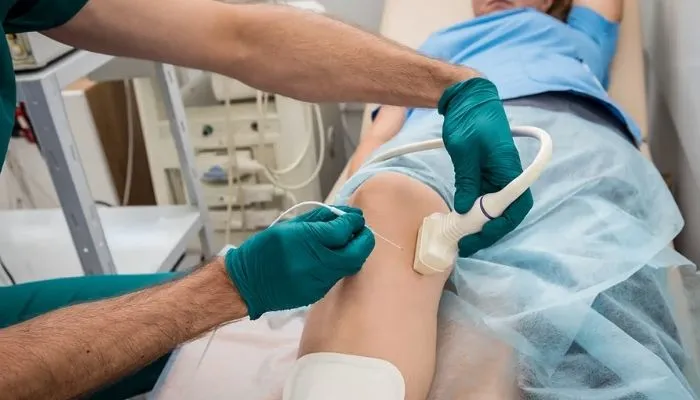
Radiofrequency Ablation (RFA) is a procedure in which a thin needle electrode is inserted through the skin directly into a tumour under ultrasound or CT image guidance. High-frequency alternating electrical current is then delivered through the electrode, causing ionic agitation in the tissue surrounding the electrode tip. This generates heat (typically 60 to 100 degrees Celsius) which destroys tumour cells through thermal coagulation necrosis - effectively cooking the tumour and killing it in place. The destroyed tissue is gradually reabsorbed by the body over the following weeks and months.
RFA is used to treat primary liver cancer (hepatocellular carcinoma), colorectal liver metastases, renal cell carcinoma (kidney cancer), lung tumours, thyroid nodules, adrenal tumours, bone metastases causing pain, and osteoid osteomas (benign bone tumours). It is particularly effective for small solid tumours (generally up to 3 to 4 cm in diameter) and for patients who are not suitable for surgery. RFA can be performed percutaneously (through the skin), laparoscopically, or as open surgery, depending on the tumour location and access requirements - at Shree Hospitals, percutaneous image-guided RFA is the primary approach.
At Shree Hospitals, RFA is performed by our interventional radiology team in dedicated procedure rooms equipped with real-time ultrasound and CT fluoroscopy. Every RFA case is discussed at the relevant multidisciplinary oncology team (MDT) meeting - hepatology MDT for liver tumours, urology MDT for renal tumours - before the procedure is recommended. Our team uses advanced internally cooled electrodes and impedance-based generators that automatically optimise energy delivery for maximum ablation zone size while minimising complications.
Who Is a Candidate for Radiofrequency Ablation?
RFA is indicated for:
- Early hepatocellular carcinoma (HCC) - solitary tumours up to 3 to 5 cm or up to 3 tumours up to 3 cm in patients not suitable for surgical resection or transplantation
- Colorectal liver metastases - limited number of metastases in patients not suitable for surgical resection
- Renal cell carcinoma - small renal tumours (under 4 cm) in patients with a single kidney, poor surgical fitness, or those wishing to preserve renal function
- Lung tumours - primary or metastatic nodules in patients not suitable for surgery
- Osteoid osteoma and painful bone metastases requiring local pain control
Is RFA the Right Treatment for Your Tumour?
RFA is most appropriate when:
- The tumour is small (generally under 3 to 4 cm for optimal complete ablation)
- The tumour is away from major blood vessels (vessels over 3mm can cause a heat sink effect reducing ablation effectiveness)
- The tumour is away from critical structures (bile ducts, bowel, ureter)
- Surgical resection is not possible due to tumour location, poor organ function, or patient co-morbidities
- The patient has adequate organ function to tolerate the procedure
- The MDT has agreed RFA is the most appropriate local treatment
Why RFA Has Transformed Tumour Treatment Options?
RFA offers significant advantages for suitable patients:
- Tumour destruction without surgery - no incision, no general anaesthesia in many cases, no surgical scar
- Organ-preserving - treats the tumour while preserving surrounding healthy liver, kidney, or lung tissue
- Repeatable - can be performed multiple times if new tumours develop
- Can be combined with TACE (for liver tumours), systemic therapy, or surgery in a multimodal approach
- Short recovery - most patients return to normal activity within 1 to 2 weeks
- Provides durable local tumour control comparable to surgery for appropriately selected small tumours
RFA achieves complete tumour destruction in over 90% of hepatocellular carcinoma tumours under 3 cm in size.
At Shree Hospitals, our interventional radiology team performs RFA using precision image guidance and advanced ablation technology, achieving durable tumour control rates comparable to surgical resection for appropriately selected small liver and kidney tumours.
24×7 Emergency Care
Our Approach to Radiofrequency Ablation
At Shree Hospitals, RFA is always preceded by careful MDT review to confirm it is the optimal local treatment for each patient's tumour. Our interventional radiologists plan every ablation using dedicated pre-procedure imaging to map the tumour, plan the electrode approach, and identify critical nearby structures that must be protected.
24×7 Emergency Care
MDT Case Review and Dedicated Pre-Procedure Planning
Every RFA candidate is reviewed at the relevant MDT meeting (liver cancer MDT, urology MDT, thoracic MDT). Triple-phase CT or contrast-enhanced MRI is used to characterise the tumour and plan the ablation. The planned ablation zone is mapped to ensure the target tumour plus a 1 cm safety margin of normal tissue is covered. Nearby critical structures - bile ducts, bowel, ureter, major blood vessels - are identified and protective manoeuvres planned where needed.
Precision Image-Guided Electrode Placement
Under CT or ultrasound guidance, the RFA electrode is advanced through the skin and positioned precisely within the centre of the target tumour. Real-time imaging confirms electrode position before ablation commences. For larger tumours, multiple overlapping ablations are performed with electrode repositioning to ensure complete coverage of the entire tumour volume plus margin.
Protective Manoeuvres for Complex Cases
When tumours are adjacent to vulnerable structures, our interventional radiologists employ protective techniques including hydrodissection (injection of saline or dextrose solution to create a buffer between the tumour and adjacent bowel or ureter), balloon displacement of adjacent bowel, and real-time temperature monitoring at the safe margin. These measures allow safe ablation of tumours that would otherwise be considered too high-risk.
Post-Ablation Imaging and Oncology Follow-Up
Contrast-enhanced CT or MRI is performed at 4 to 6 weeks after RFA to confirm complete tumour ablation (absence of any residual enhancement at the ablation site). Response is assessed using mRECIST criteria. Residual or recurrent tumour is discussed at the MDT and managed with repeat ablation, TACE, or transition to systemic therapy. Long-term surveillance imaging continues at regular intervals per oncology guidelines.
Interventional Radiologists and Oncology Specialists in Mumbai
Book Expert Consultation

10+ years of experience
Dr. Swati Narurkar
Interventional Radiology
Interventional Radiology Services at Shree Hospitals
We offer a comprehensive range of minimally invasive interventional radiology procedures including radiofrequency ablation, TACE, UAE, percutaneous drainage, biliary drainage, peripheral angioplasty, vertebroplasty, and IVC filter placement, all delivered by specialist interventional radiologists using advanced imaging guidance.
Step by Step Process of Radiofrequency Ablation
Step 1 - MDT Review and Pre-Procedure Assessment
Your case is presented at the multidisciplinary oncology team meeting. Contrast-enhanced imaging of the tumour is reviewed and the ablation plan is designed. Blood tests including liver function, kidney function, and clotting profile are checked. Medical fitness for conscious sedation or general anaesthesia is assessed. The procedure, expected outcomes, and potential complications are explained and consent is obtained.
Step 2 - Patient Preparation and Anaesthesia
RFA is performed under general anaesthesia or deep conscious sedation depending on tumour location, expected procedure duration, and patient factors. Intravenous access is established and pre-operative medications given. For kidney tumours, ureteric stents may be placed by urology in advance if the tumour is near the ureter. The patient is positioned for optimal imaging and access.
Step 3 - Image-Guided Electrode Placement
Using real-time CT or ultrasound guidance, the RFA electrode needle is advanced through the skin via a carefully planned trajectory to reach the centre of the target tumour. A targeting CT scan confirms precise electrode position within the tumour before ablation is started. The electrode tip is confirmed to be within the tumour mass with an adequate margin.
Step 4 - Radiofrequency Ablation
The RFA generator is activated and radiofrequency energy is delivered through the electrode. Energy output, tissue impedance, and temperature are monitored in real-time. The ablation is continued until the target zone diameter (tumour plus 1 cm margin) is achieved - typically 10 to 15 minutes per ablation position. The electrode is repositioned for overlapping ablations in larger tumours. Ablation completion is confirmed by the characteristic change in tissue impedance.
Step 5 - Track Ablation and Electrode Withdrawal
As the electrode is withdrawn, the electrode track is ablated to prevent tumour seeding along the needle path - a technique known as track cauterisation. A post-ablation CT scan is performed immediately to confirm the ablation zone, check for complications (bleeding, pneumothorax for lung ablations), and document the initial result.
Step 6 - Recovery and Oncology Follow-Up
Most patients are monitored for 24 to 48 hours after RFA. Post-ablation fever and discomfort (post-ablation syndrome) are managed with analgesia and anti-pyretics. Most patients are discharged within 1 to 2 days and return to normal activity within 1 to 2 weeks. Contrast-enhanced CT or MRI at 4 to 6 weeks confirms complete ablation. Long-term follow-up imaging is continued per the relevant oncology surveillance protocol.
Patient Questions About Radiofrequency Ablation
Is RFA as effective as surgery for liver tumours?
For small liver tumours (under 3 cm), RFA achieves complete ablation rates comparable to surgical resection with similar long-term survival outcomes in appropriately selected patients. Studies show no significant difference in 5-year survival between RFA and surgical resection for HCC tumours under 2 cm. For larger tumours (3 to 5 cm), surgery may achieve better margins but RFA remains an important alternative for patients who cannot undergo surgery. The best treatment for each patient is determined by the MDT based on tumour characteristics, liver function, and overall health
Can RFA be repeated if the tumour comes back?
Yes. RFA can be repeated if a treated tumour shows local recurrence at the ablation margin, or if new tumours develop elsewhere in the same organ. This is one of the significant advantages of RFA over surgery - it can be performed multiple times without the cumulative risks of repeated surgical resection. However, liver function must be adequate for each session. Between sessions, oncology surveillance imaging monitors for recurrence. Recurrent tumours are discussed at the MDT to determine whether repeat RFA, TACE, or systemic therapy is most appropriate.
What organs can be treated with RFA?
RFA at Shree Hospitals can treat tumours in the liver (most common), kidney, lung, adrenal glands, thyroid gland (for benign nodules and selected small cancers), and bone (painful metastases and osteoid osteoma). The specific application depends on the tumour type, size, and location relative to critical structures. Thyroid RFA is a growing application for treating benign thyroid nodules causing pressure symptoms or cosmetic concerns without surgery. Each case is reviewed by the relevant MDT to confirm the appropriateness of RFA for the specific tumour.
How do I know if the RFA worked?
The success of RFA is assessed by contrast-enhanced imaging (CT or MRI) performed at 4 to 6 weeks after the procedure. A complete response is defined as no enhancement (no blood flow) within the ablation zone on imaging, indicating complete tumour destruction. If any residual viable tumour (areas of enhancement) is seen at the margin, repeat ablation can be performed. After confirmed complete response, regular surveillance imaging continues to detect any local recurrence or new tumours early.
Patient
Resources

Schedule
consultation

Billing &
Payments

Hospital
Facilities
24×7 Emergency Care

1800-268-4000

Follow Us

Shree
Hospitals

Shree
Hospitals
