 Ghatkopar (East), Mumbai)

Home > Neurosurgery > Complex Spinal Reconstruction
Complex Spinal Reconstruction In Mumbai
Osteotomy, Long-Segment Instrumented Fusion, Spinal Tumour Resection, and Sagittal Balance Correction at Shree Hospitals
Complex spinal reconstruction covers the most technically demanding spine surgery - procedures requiring multilevel instrumented fusion, corrective osteotomies for severe spinal deformity, spinal tumour resection (including intradural and intramedullary spinal tumours), management of spinal infection with instability, and correction of failed previous spinal surgery. At Shree Hospitals, our complex spinal reconstruction programme brings together specialist neurosurgeons and orthopaedic spine surgeons, neurophysiologists for continuous intraoperative monitoring, oncologists and radiation oncologists for spinal tumour management, and vascular and general surgeons where anterior surgical approaches require it. We perform the full range of reconstructive procedures: Smith-Petersen osteotomy (SPO), Ponte osteotomy, pedicle subtraction osteotomy (PSO), vertebral column resection (VCR), long-segment instrumented fusion with pedicle screws, rods, and interbody cages, corpectomy with expandable cage reconstruction, and en bloc resection of spinal tumours classified using the Weinstein-Boriani-Biagini (WBB) system.
Experiencing persistent neurological symptoms? Schedule a consultation with our expert neurosurgeons today.
Quick facts
Procedures: SPO, PSO, VCR, Long-Segment Fusion, Corpectomy, En Bloc Tumour Resection
Conditions treated: Scoliosis, Kyphosis, Spinal Tumour, Spinal Infection, Failed Spinal Surgery
Approach: Posterior, Anterior, or Combined (360-degree)
Anaesthesia: General Anaesthesia with Continuous Intraoperative Neurophysiological Monitoring
Hospital stay: 5 to 14 days, depending on procedure complexity
Key technology: O-arm Navigation, IONM (SSEP/MEP/EMG), Expandable Cages, Neuromonitoring
What is Complex Spinal Reconstruction and Who Needs It?
Complex spinal reconstruction is a broad surgical category encompassing procedures that go beyond routine discectomy or single-level fusion - operations that require multilevel instrumentation, correction of fixed spinal deformity, resection of spinal tumours, or reconstruction after spinal infection or previous failed surgery. The hallmark of complex spinal reconstruction is the requirement to restore global spinal alignment (the balance of the spine in both the sagittal and coronal planes), decompress the spinal cord or nerve roots, achieve long-term spinal stability, and (in tumour and infection cases) eradicate pathological tissue. The most common indications are: adult and adolescent spinal deformity (scoliosis with Cobb angle above 45 to 50 degrees requiring surgery, and kyphosis with fixed sagittal imbalance); spinal tumours (primary bone tumours, spinal metastases, and intradural tumours); spinal infection with vertebral collapse and instability (discitis, osteomyelitis, tuberculosis of the spine - Pott's disease); and failed spinal surgery requiring revision and reconstruction.
The key concept in complex spinal reconstruction - especially deformity correction - is sagittal balance. The spine is designed to maintain the head over the pelvis in the most energy-efficient posture. Sagittal vertical axis (SVA) is the horizontal distance between a vertical line dropped from the C7 vertebra and the back of the sacrum - in a normally aligned spine this is under 50mm. When the spine develops fixed deformity (rigid kyphosis, failed flat back after previous lumbar fusion, progressive scoliosis with coronal collapse), the SVA increases and patients adopt compensatory mechanisms (hip extension, knee flexion, pelvic retroversion) that are exhausting and painful. Corrective osteotomies are the surgical tools to restore SVA: SPO (opening wedge through the posterior elements, 5 to 10 degree correction per level), Ponte osteotomy (posterior column shortening, 10 to 15 degree correction), PSO (all three columns included, 30 to 40 degree correction per level), and VCR (complete vertebral column resection, up to 120 degree correction for the most severe deformities). The choice of osteotomy is guided by the rigidity and magnitude of the deformity, the number of levels requiring correction, and the medical fitness of the patient for a prolonged, high-blood-loss surgical procedure.
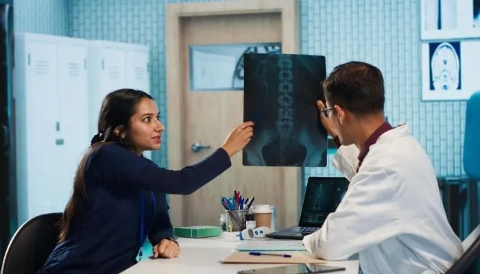
At Shree Hospitals, complex spinal reconstruction cases are planned and executed by a multidisciplinary spine team with representatives from neurosurgery, orthopaedic spine surgery, anaesthesia, neurophysiology, oncology (for tumour cases), and intensive care. Pre-operative planning is meticulous: standing full-spine X-rays with measurement of spinal alignment parameters (SVA, PI, LL, TK, C7 plumb line, pelvic incidence-lumbar lordosis mismatch), CT for bony anatomy and pedicle dimensions, MRI for cord and root compression, tumour characteristics, and infection extent, and bone density assessment (DEXA) for implant planning. Intraoperative neurophysiological monitoring (IONM) with simultaneous SSEP, MEP, and free-running and triggered EMG is mandatory for all complex reconstruction procedures.
Who Needs Complex Spinal Reconstruction?
Complex spinal reconstruction is indicated for:
- Adult scoliosis or kyphosis with Cobb angle above 45 to 50 degrees, progressive deformity causing pain and functional disability, or neurological compromise from cord or root compression from the deformity
- Fixed sagittal imbalance (flat back syndrome after previous lumbar fusion, iatrogenic flatback from long-segment fusion without adequate lordosis restoration) causing severe disability - unable to look horizontal without bending the knees
- Spinal metastases causing vertebral body collapse with cord compression (malignant spinal cord compression - MSCC) requiring surgical decompression and stabilisation before radiotherapy, or single-level solitary metastasis in good-prognosis cancer where separation surgery or en bloc resection is appropriate
- Primary spinal tumours - benign (osteoid osteoma, osteoblastoma, giant cell tumour) and malignant (chordoma, osteosarcoma, Ewing's sarcoma, plasmacytoma) requiring en bloc resection with WBB classification planning
- Intradural spinal tumours - extramedullary (meningioma, schwannoma, neurofibroma) and intramedullary (ependymoma, astrocytoma, haemangioblastoma) requiring microsurgical resection under IONM
- Pott's disease (spinal tuberculosis) with kyphotic deformity, vertebral collapse, and cord compression - requiring anterior debridement, fusion, and posterior instrumented stabilisation
What are Osteotomies and How Do They Correct Spinal Deformity?
Osteotomies are deliberate surgical cuts through the bone and posterior elements of the spine to allow correction of rigid deformity:
- Smith-Petersen Osteotomy (SPO): The posterior elements (spinous process, laminae, and facet joints) are resected at one level, creating an opening wedge. The spine is then extended to close the wedge and gain lordosis. Requires that the anterior disc space is mobile enough to open anteriorly as the posterior elements close. Correction: approximately 5 to 10 degrees per level. Multiple levels can be performed simultaneously
- Ponte Osteotomy: Similar to SPO but used in the thoracic spine for kyphosis correction. The posterior elements are resected bilaterally and the spine is compressed posteriorly. Used for Scheuermann's kyphosis and ankylosing spondylitis. Correction: approximately 10 to 15 degrees per level
- Pedicle Subtraction Osteotomy (PSO): All three columns of the spine are included - the posterior elements and the pedicles are resected, and a closing wedge is taken from the posterior two-thirds of the vertebral body, allowing the posterior elements to close and the vertebral body to close like a wedge. The anterior column is not disrupted. Correction: 30 to 40 degrees per single level - the most powerful single-level correction that does not require anterior surgery
- Vertebral Column Resection (VCR): The entire vertebral body (or multiple vertebral bodies), the adjacent discs, and all posterior elements at those levels are resected. The spinal cord or cauda equina is freed circumferentially. The two ends of the spine are then brought together and stabilised with a cage anteriorly and long-segment pedicle screw instrumentation posteriorly. Correction: up to 120 degrees for the most severe angular deformities, including sharp post-traumatic or congenital kyphosis
How Spinal Tumours are Classified and Planned for Surgery
The Weinstein-Boriani-Biagini (WBB) classification is the internationally accepted system for planning the surgical approach and extent of resection for primary spinal tumours with curative intent:
- The vertebra is divided into 12 sectors around the circumference (like a clock face) and 5 layers from the paravertebral soft tissue to the epidural space, allowing precise mapping of tumour extent
- Marginal or wide resection (en bloc) is the goal for primary malignant spinal tumours - removing the entire tumour in one piece with a margin of normal tissue, without entering the tumour (intralesional resection). This is the same oncological principle as en bloc resection of bone sarcomas in the limbs, applied to the spine
- The challenge of en bloc spinal resection is the proximity of the spinal cord - the surgeon must resect the tumour in one piece while carefully protecting the cord throughout, which requires meticulous planning, often staged anterior then posterior surgery, and extensive vascular control
- Intradural extramedullary tumours (meningioma, schwannoma) are resected using microsurgery under IONM - schwannomas are generally completely resectable with excellent outcomes (recurrence under 5%); meningiomas are resected with their dural attachment
- Intramedullary tumours (within the spinal cord itself - ependymoma, astrocytoma) require the highest level of microsurgical skill and IONM - ependymomas are often resectable from within the cord using the midline myelotomy approach; astrocytomas may be only partially resectable
Complex Spinal Reconstruction at Shree Hospitals restores spinal alignment, decompresses the spinal cord, and achieves long-term stability in the most challenging spinal conditions - scoliosis, tumour, infection, and failed surgery.
Our multidisciplinary complex spine team at Shree Hospitals uses O-arm navigation, continuous intraoperative SSEP, MEP, and EMG monitoring, corrective osteotomies (SPO, Ponte, PSO, VCR), expandable titanium cage reconstruction, and en bloc tumour resection planning with WBB classification to deliver safe and effective complex spinal reconstruction with the highest standards of neurological protection.
24×7 Emergency Care
Our Approach to Complex Spinal Reconstruction at Shree Hospitals
Complex spinal reconstruction at Shree Hospitals follows a structured approach: meticulous pre-operative planning with full-length standing spinal imaging, multidisciplinary team planning for tumour and infection cases, surgical execution with continuous neurological monitoring, and structured post-operative rehabilitation.
24×7 Emergency Care
Comprehensive Pre-Operative Assessment and Planning
Every complex spinal reconstruction case begins with a formal pre-operative planning meeting attended by the spine surgeon, neurophysiologist, anaesthetist, and (for tumour cases) oncologist. Imaging is reviewed in full: standing full-spine EOS or full-length X-rays with measurement of all global alignment parameters (SVA, C2-C7 plumb, T1 slope, TK, LL, SS, PI, and PI-LL mismatch); MRI for neural compression, cord signal change, tumour or infection extent, and intradural detail; CT for pedicle morphology, bone quality, and tumour/infection bony involvement; and bone density (DEXA) where osteoporosis is suspected. For spinal tumours, additional PET-CT or bone scan is obtained to assess for systemic disease. 3D CT reconstruction is used for osteotomy simulation and pedicle screw trajectory planning. Blood transfusion planning includes pre-operative haematinics, cell salvage, and normovolaemic haemodilution for long-segment deformity procedures (blood loss can be 2 to 5 litres in major deformity surgery).
Long-Segment Instrumented Fusion with Corrective Osteotomy
For adult deformity correction, the surgical approach begins with the patient prone on a radiolucent Jackson table. Posterior exposure is performed (open approach for complex deformity; hybrid approaches with MIS components where possible). Pedicle screws are inserted at all levels to be fused - typically from T2 or T3 to the ilium for long thoracolumbar fusions - under navigation guidance to ensure accurate screw placement in potentially dysplastic or rotated pedicles. The corrective osteotomy is then performed at the apex of the deformity (PSO for sagittal plane correction, or combined SPO/Ponte for thoracic kyphosis). Cantilever reduction is performed against the rod to gradually close the osteotomy and correct the deformity. IONM is monitored continuously throughout - any significant change in SSEP amplitude or MEP disappearance results in immediate reversal of correction and investigation. After deformity correction, interbody fusion cages are inserted at the osteotomy level to restore anterior column support.
Spinal Tumour Resection and Reconstruction
For malignant spinal cord compression (MSCC) from metastatic disease, the most common presentation, the goal is surgical decompression of the spinal cord (removing the tumour compressing the cord) and stabilisation of the spine to allow subsequent radiotherapy. This is typically achieved through a posterior approach with laminectomy, pedicle resection, and posterolateral decompression combined with pedicle screw fixation above and below the involved levels - a procedure that can be performed relatively quickly (2 to 4 hours). For primary malignant spinal tumours, en bloc resection using WBB classification planning is performed - often staged (anterior then posterior or vice versa) over 1 to 2 days. After tumour resection, vertebral body reconstruction uses expandable titanium mesh cages or 3D-printed titanium cages for anterior column support, combined with long-segment posterior instrumentation. For intradural tumours, microsurgery is performed through a laminectomy approach with the operating microscope and IONM throughout. Sharp microsurgical dissection and ultrasonic aspirator (CUSA) are used for intramedullary tumour debulking.
Spinal Infection Management
Spinal infection (discitis, osteomyelitis, Pott's disease - spinal tuberculosis) with cord or nerve root compression, vertebral collapse, or progressive kyphotic deformity requires surgical treatment. The principles are: radical debridement of infected and necrotic tissue (anterior approach for anterior column infection), reconstruction of the anterior column with structural bone graft or expandable cage (titanium mesh is generally avoided in active infection in favour of structural autograft - typically rib or fibula), and posterior instrumented stabilisation to provide immediate stability while the anterior reconstruction incorporates. The posterior instrumentation is placed outside the zone of infection where possible (hooks or screws above and below the infected levels). For Pott's disease (TB spine), surgery is combined with a 9 to 12 month antituberculous therapy regimen. The correction of the Pott's kyphosis (Gibbus deformity) if severe may require osteotomy at the same time as debridement, or as a staged procedure once infection is controlled.
Top Neurosurgeons in Mumbai
Every specialist at Shree Hospitals is MCh Neurosurgery board-certified with over 15 years of clinical experience in awake craniotomy, brain tumour surgery, neuronavigation, fluorescence-guided resection, functional brain mapping, and neuro-oncology.
24×7 Emergency Care

Dr. Yashwant Shelke
DNB Neurosurgery
Dr. Survendra Rai
Micro-Neurosurgery, Minimally Invasive Spine Surgery

Dr. Medha Vyas
Consultant Neurosurgeon & NeuroSpine Surgeon
Dr. Viswanathan Iyer
Consultant Neurosurgeon & Endovascular
Neurosurgery Services at Shree Hospitals
We provide a comprehensive range of neurosurgery and spine surgery services including complex spinal reconstruction, minimally invasive spine surgery, stereotactic procedures, Gamma Knife radiosurgery, and awake craniotomy.
Step by Step Process of Complex Spinal Reconstruction at Shree Hospitals
Step 1 - Multidisciplinary Team Assessment
The patient is reviewed at the complex spine MDT meeting with all relevant specialties. Imaging is presented, the surgical indication is confirmed, and the surgical plan is agreed: the type and number of osteotomies, the levels to be instrumented (screws/hooks above and below), the anterior column reconstruction strategy (interbody cage type and size, bone graft source), the surgical staging (single or two-stage procedure), and the approach (posterior alone, anterior-posterior, lateral). Oncology input is obtained for spinal tumour cases (systemic staging, chemotherapy status, expected prognosis - critical for deciding the extent of reconstruction in metastatic disease). Blood transfusion service is involved for major deformity cases.
Step 2 - Pre-Operative Preparation
Pre-operative haematinics (iron therapy, EPO if indicated) are optimised to maximise pre-operative haemoglobin. Cell salvage (intraoperative blood salvage and reinfusion) is arranged for all major deformity procedures. IONM electrodes are placed by the neurophysiologist after anaesthetic induction. Positioning is on a Jackson radiolucent table (prone) or lateral decubitus (for anterior or lateral approaches). Position is confirmed with fluoroscopy. O-arm intraoperative CT is acquired for navigation registration.
Step 3 - Posterior Instrumentation
Through a posterior midline incision, the spine is exposed at all levels to be instrumented. Pedicle screws are inserted under navigation guidance at each instrumented level. In deformity cases with pedicle dysplasia or rotation (common in scoliosis), navigation-guided screw placement is essential for accuracy. Pedicle probing and triggered EMG confirm each screw is safely within the pedicle cortex. Provisional rods are placed to maintain alignment while osteotomies are performed.
Step 4 - Corrective Osteotomy
The osteotomy is performed at the planned level(s). For PSO: the spinous process, lamina, transverse processes, and pedicles at the osteotomy level are resected. A closing wedge is cut into the posterior and middle vertebral body, leaving the anterior cortex intact as the hinge. For VCR: complete resection of the vertebral body and all posterior elements, with the cord protected throughout by careful bipolar coagulation and dissection. For SPO/Ponte: posterior element resection only, relying on disc mobility anteriorly. After osteotomy, cantilever reduction against the rods gradually closes the osteotomy and corrects the deformity. IONM is monitored in real-time throughout this phase - the highest-risk phase for neurological injury.
Step 5 - Anterior Column Reconstruction
For corpectomy and vertebrectomy cases (tumour, infection, VCR), the anterior column must be reconstructed. An expandable titanium mesh cage or 3D-printed titanium cage is sized to the corpectomy defect and packed with bone graft material (autograft from the resected posterior elements, or allograft). The cage is inserted into the corpectomy defect and expanded to achieve a press-fit reconstruction, restoring anterior column height and lordosis. In tumour cases, the cage is sized to span at least one level above and below the vertebrectomy to prevent endplate subsidence.
Step 6 - Final Fixation, IONM Confirmation, and Closure
Final rod contouring and set-screw tightening completes the posterior instrumentation construct. An O-arm spin is acquired for post-reduction imaging - confirming the deformity correction, the interbody cage position, and all screw positions in the corrected anatomy. IONM is confirmed stable throughout. The wound is closed in layers over suction drains. The patient is transferred to the neurosurgical intensive care unit (ICU) for the first 24 to 48 hours for neurological monitoring and haemodynamic management. Post-operative standing X-rays at 6 weeks assess the achieved correction and early fusion progress.
Patient Questions About Complex Spinal Reconstruction
How long does complex spinal reconstruction surgery take and what is the blood loss?
Major complex spinal reconstruction is among the longest and most demanding of all surgical procedures. A long-segment adult scoliosis correction from the thoracic spine to the ilium with one or two PSOs typically takes 8 to 14 hours of operating time. Blood loss can be 2 to 5 litres - equivalent to the patient's total blood volume in major VCR cases. This is managed with: pre-operative blood donation (if time allows), pre-operative haematinics and EPO to maximise pre-operative haemoglobin, intraoperative cell salvage (blood lost is collected, processed, and reinfused), tranexamic acid (an antifibrinolytic that significantly reduces blood loss in major spine surgery), and blood transfusion where required. The anaesthetic team at Shree Hospitals for major deformity cases is highly experienced in managing the haemodynamic challenges of prolonged major spine surgery. Patients should expect to spend at least one night in the ICU for intensive monitoring after major reconstruction.
What is the risk of paralysis in complex spinal reconstruction?
Neurological injury - from numbness and weakness to, in the most severe cases, permanent paralysis - is the most serious complication of complex spinal reconstruction and is a real and unavoidable risk that all patients must understand before consenting to surgery. At Shree Hospitals, we minimise this risk with continuous IONM (SSEP + MEP + EMG) throughout all complex reconstruction procedures. If the neurophysiologist reports a significant change in SSEP amplitude or the loss of MEPs during the correction manoeuvre, the surgical team immediately responds: reversing the correction, checking for mechanical cord compression, raising blood pressure to improve cord perfusion, and administering steroids. The "wake-up test" (asking the patient to move their feet during surgery under lighter anaesthesia) can be performed as an additional neurological check during major correction.
How long is the recovery after complex spinal reconstruction?
Recovery after complex spinal reconstruction is prolonged - patients should plan for a 6 to 12 month recovery period for major procedures. In hospital, physiotherapy begins on post-operative day 1 or 2 (earlier for decompression cases, later for major deformity correction). Most patients are mobilised with a thoracolumbar spinal orthosis (TLSO brace) for 3 to 6 months after major deformity surgery to protect the instrumentation while the fusion consolidates. The bone fusion across the operated levels takes 3 to 6 months to become solid radiologically, and up to 12 to 18 months for full maturation.
Can spinal metastases be treated surgically at Shree Hospitals?
Yes - Shree Hospitals offers the full range of surgical treatment for spinal metastases, from minimally invasive decompression and stabilisation for acute cord compression through to en bloc resection for selected cases. The decision on the extent of surgery for metastatic spinal disease is made by the oncology MDT and takes into account the primary tumour histology and prognosis (using tools such as the Tokuhashi score and Tomita score), the degree of cord compression and neurological deficit, the stability of the spine, the expected response to radiotherapy, the number of spinal levels involved, and the patient's fitness for surgery.

Evidence-Based Case Studies by Our Specialists
Would Recommend Us
Adult scoliosis with 72-degree Cobb angle and severe sagittal imbalance (SVA 110mm) in a 58-year-old with progressive back pain and inability to stand upright, treated with T3 to ilium posterior instrumented fusion with bilateral PSO at L3, achieving Cobb correction to 28 degrees and SVA correction to 32mm. Patient discharged day 8, returned to normal daily activities at 4 months.
Mr. Kisan
L1 spinal metastasis from renal cell carcinoma with complete L1 vertebral body destruction and early cord compression in a 62-year-old with good systemic disease control, treated with T11 to L3 posterior instrumented stabilisation and L1 corpectomy with expandable cage reconstruction, followed by SBRT (24Gy in 2 fractions) to the L1 level. Ambulatory at discharge, full weight-bearing at 2 weeks.
Mr. Ravi
Thoracic spinal tuberculosis (Pott's disease) at T7/T8 with 35-degree kyphosis, cord compression, and early weakness of both legs (ASIA Grade D) in a 36-year-old, treated with T5 to T10 posterior instrumented stabilisation followed by anterior debridement, structural rib graft, and partial kyphosis correction (to 18 degrees). Antituberculous therapy for 12 months. Neurological recovery to ASIA Grade E at 6 months, returned to work at 8 months.
Mr. Deva
Patient
Resources

Schedule consultation

Billing & Payments

Health Packages

Hospital Facilities
24×7 Emergency Care

1800-268-4000

News & Updates
Shree Hospitals is the first to perform robotic endo surgery.
Shree Hospitals Introduces Advanced Robotic Surgery for Better Patient Care
Shree Hospitals Expands Advanced Surgical Care With Latest Medical Technology
Health Center
Exploring the Future of Telemedicine: What’s Next for Healthcare?
The Rise of Renewable Energy: Innovations in Sustainability
Follow Us

Shree
Hospitals

Shree
Hospitals
