 Ghatkopar (East), Mumbai)

Home > Neurosurgery > Stereotactic Procedures
Stereotactic Procedures In Mumbai
Expert Stereotactic Brain Biopsy and Deep Brain Stimulation (DBS) for Parkinson's Disease, Essential Tremor, and Dystonia at Shree Hospitals
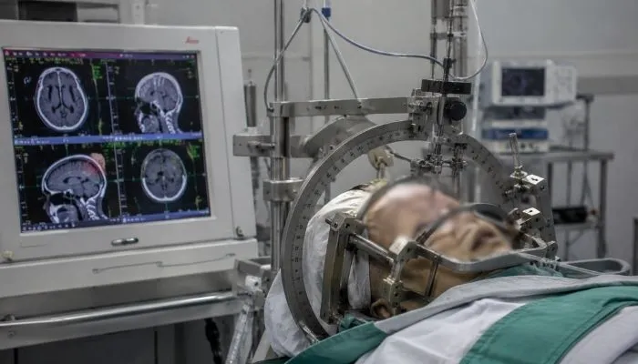
Stereotactic procedures use a precise three-dimensional coordinate system - derived from brain imaging (MRI and CT) - to guide surgical instruments to specific targets deep within the brain with millimetre accuracy, without the need for wide open craniotomy. At Shree Hospitals, our specialist neurosurgeons perform two primary categories of stereotactic procedures: stereotactic brain biopsy - obtaining tissue samples from deep-seated or eloquent-region brain lesions where open surgery is not safe, providing the diagnosis that guides treatment; and deep brain stimulation (DBS) - implanting electrodes into precisely targeted deep brain nuclei to treat Parkinson's disease, essential tremor, and dystonia - the most effective surgical treatment for movement disorders.
Experiencing persistent neurological symptoms? Schedule a consultation with our expert neurosurgeons today.
Quick facts
Stereotactic biopsy: Deep-seated Brain Tumour, Inflammatory Lesion, Infectious Lesion, Tissue Diagnosis
DBS targets: STN (Parkinson's), VIM (Tremor), GPi (Dystonia, Parkinson's)
DBS indications: Parkinson's Disease, Essential Tremor, Dystonia, OCD (selected cases)
Anaesthesia: Biopsy: Local/GA; DBS: Awake (for electrophysiology), then GA for IPG
Hospital stay: Biopsy: 1 to 2 days; DBS: 2 to 3 days (two-stage procedure)
What are Stereotactic Neurosurgical Procedures?
Stereotaxis (from the Greek "stereos" - three-dimensional, and "taxis" - arrangement) refers to the use of a three-dimensional coordinate system to locate a target within the brain precisely from imaging data. By registering a patient's brain MRI or CT to a stereotactic frame (or frameless neuronavigation system), every point within the brain can be assigned exact x, y, and z coordinates. A surgical instrument - biopsy needle, electrode, or laser fibre - can then be guided to any target point along a pre-planned trajectory, passing through the safest possible corridor of brain tissue (avoiding eloquent cortex, blood vessels, and ventricles) with millimetre accuracy.
Stereotactic brain biopsy uses this system to obtain a small tissue sample (1 to 3mm cores) from a deep-seated or eloquent brain lesion that cannot be safely approached by open surgery. Common targets include: deep-seated gliomas in the thalamus, basal ganglia, or brainstem; CNS lymphoma (which is exquisitely sensitive to chemotherapy but must be distinguished histologically from other tumours); inflammatory or demyelinating lesions (MS, CNS vasculitis, sarcoidosis); infectious lesions (cerebral abscess, tuberculoma, toxoplasmosis); and any brain lesion where the tissue diagnosis will change management but open resection is not indicated.
Deep brain stimulation (DBS) is a neurosurgical procedure in which thin, multi-contact electrodes are implanted into specific deep brain nuclei and connected to an implantable pulse generator (IPG) - a programmable pacemaker-like device implanted under the skin of the chest wall or abdomen. Electrical stimulation delivered through the electrodes at specific parameters modulates the abnormal firing patterns of the target nucleus, suppressing the motor symptoms of Parkinson's disease (tremor, rigidity, bradykinesia, on-off fluctuations), essential tremor (disabling hand tremor), and dystonia (sustained abnormal postures and involuntary movements).
Who Needs a Stereotactic Procedure?
Stereotactic biopsy is indicated for:
- Any deep-seated brain lesion (thalamus, basal ganglia, brainstem, corpus callosum, internal capsule) that cannot be safely resected but needs histological diagnosis
- CNS lymphoma - strongly suspected on MRI (homogeneous enhancement, periventricular, restricted diffusion) but must be biopsied before any treatment including steroids (steroids can temporarily eliminate lymphoma, causing a false-negative biopsy)
- Multiple small ring-enhancing lesions - differential includes metastases, lymphoma, infection, demyelination - biopsy resolves the diagnosis
- Radiological diagnosis uncertainty - where the MRI appearance does not permit confident clinical diagnosis
DBS is indicated for:
- Parkinson's disease: motor fluctuations and dyskinesias despite optimised dopaminergic therapy, with preserved cognitive function and good response to levodopa
- Essential tremor: disabling hand tremor not controlled by propranolol, primidone, or other medications
- Dystonia: generalised or focal dystonia causing significant disability
Is a Stereotactic Procedure Right for You?
Stereotactic biopsy is the right choice when:
- The tissue diagnosis is essential for treatment planning (you cannot treat a brain tumour without knowing its type)
- Open resection is not safe or not indicated for the lesion (e.g., diffuse brainstem glioma, thalamic tumour)
- Steroids have not yet been given (for suspected CNS lymphoma)
DBS is the right choice for Parkinson's when:
- You have had Parkinson's disease for 5 or more years and medications are no longer providing adequate control
- You have a good response to levodopa (the therapeutic response test confirms DBS suitability - if levodopa does not significantly improve your symptoms, DBS is unlikely to help)
- You have preserved cognition on neuropsychological testing (DBS is not recommended for patients with significant dementia)
- You do not have significant depression or other psychiatric comorbidity that DBS might worsen
- Your movement disorder neurologist has confirmed DBS candidacy after thorough assessment
Why DBS Is the Most Effective Treatment for Advanced Parkinson's Disease?
Deep brain stimulation achieves what medications cannot in advanced Parkinson's disease:
- Continuous 24-hour symptom control: Parkinson's medications wear off between doses, causing predictable on-off fluctuations that severely affect quality of life. DBS provides continuous stimulation without the peaks and troughs of tablet-based therapy
- Reduction in levodopa dose: DBS reduces the total daily levodopa dose by 40 to 60% on average, reducing medication-induced dyskinesias
- Motor benefits: STN-DBS achieves 50 to 70% reduction in UPDRS III motor score (the standard Parkinson's disability assessment) in the medication-off state
- Quality of life: patients report dramatic improvements in ability to walk, dress, and perform activities of daily living - transforming independence
- Adjustability: DBS stimulation parameters can be adjusted non-invasively at follow-up visits (and remotely in modern systems) to optimise the benefit as the disease progresses
Stereotactic DBS at Shree Hospitals delivers 50 to 70% reduction in Parkinson's motor symptoms, restoring independence and transforming quality of life.
Our neurosurgery and movement disorder team at Shree Hospitals uses MRI-guided stereotactic targeting with intraoperative microelectrode recording (MER) for precise STN, GPi, and VIM nucleus localisation, delivering DBS electrode placement with millimetre accuracy and a comprehensive post-operative stimulation programming service to optimise outcomes for every patient.
24×7 Emergency Care
Our Approach to Stereotactic Procedures at Shree Hospitals
At Shree Hospitals, stereotactic brain biopsy and DBS surgery are performed by a specialist team that combines neurosurgical precision with advanced intraoperative imaging and neurophysiology to achieve the best diagnostic and therapeutic outcomes.
24×7 Emergency Care
Stereotactic Brain Biopsy
Planning and Trajectory Selection For stereotactic brain biopsy, the key to diagnostic success and safety is meticulous trajectory planning. The target is identified on the pre-operative MRI and mapped in the stereotactic coordinate system. A biopsy trajectory (the angle of approach of the biopsy needle) is then selected that passes through the safest corridor of brain tissue: avoiding the eloquent cortex, the corticospinal tract (motor tract), major sulci and vessels, the lateral ventricles, and the deep venous structures. For enhancing lesions, the biopsy target is placed at the most enhancing point (highest cellularity tumour).
Stereotactic Brain Biopsy
Technique and Safety Stereotactic biopsy is performed under local anaesthesia (scalp and burr hole site) with mild sedation, or under general anaesthesia in patients who cannot cooperate or where the target location requires a more complex approach. A burr hole (1.5 to 2cm circular opening) is made with a twist drill through the skull at the planned entry point. The biopsy cannula is advanced along the stereotactic arc to the target depth. Multiple cores are taken with a side-cutting biopsy needle (Sedan or similar).
DBS Surgery
Target Selection and Electrode Placement For DBS, target selection is diagnosis-dependent: the subthalamic nucleus (STN) is the primary target for Parkinson's disease (best overall motor benefit and greatest reduction in medication requirement); the ventral intermediate nucleus of the thalamus (VIM) is the preferred target for essential tremor (immediate and dramatic tremor suppression); the globus pallidus internus (GPi) is used for Parkinson's with prominent dyskinesias and for dystonia.
DBS Post-Operative Programming and Long-Term Management
DBS is not "one operation and done" - post-operative stimulation programming is as important as the surgical placement. After implantation, the DBS system is activated 2 to 4 weeks post-operatively. An initial programming session sets the active electrode contact, pulse width, frequency, and amplitude to maximise therapeutic benefit. Multiple programming sessions over the following months fine-tune the parameters. STN DBS programming is performed in the off-medication state to isolate the stimulation effect from the medication effect.
Top Neurosurgeons in Mumbai
Every specialist at Shree Hospitals is MCh Neurosurgery board-certified with over 15 years of clinical experience in awake craniotomy, brain tumour surgery, neuronavigation, fluorescence-guided resection, functional brain mapping, and neuro-oncology.
24×7 Emergency Care

Dr. Yashwant Shelke
DNB Neurosurgery

Dr. Survendra Rai
Micro-Neurosurgery, Minimally Invasive Spine Surgery

Dr. Medha Vyas
Consultant Neurosurgeon & NeuroSpine Surgeon

Dr. Viswanathan Iyer
Consultant Neurosurgeon & Endovascular
Neurosurgery Services at Shree Hospitals
We offer a comprehensive range of neurosurgery procedures including stereotactic biopsy, deep brain stimulation, awake craniotomy, Gamma Knife radiosurgery, minimally invasive spine surgery, and endoscopic skull base surgery.
Step by Step Process of Stereotactic Procedures at Shree Hospitals
Step 1 - Pre-Operative Planning and MDT Review
For biopsy: the brain lesion is reviewed at the Neuro-Oncology MDT to confirm stereotactic biopsy is the appropriate approach (not open resection) and to plan the imaging protocol for biopsy planning (gadolinium MRI, MRS, perfusion MRI). For DBS: the patient is assessed by the movement disorder neurologist (UPDRS III on and off medication, neuropsychological testing, psychiatric assessment, MRI to exclude structural abnormality), and the DBS candidacy is confirmed at the Movement Disorder MDT. The pre-operative MRI for DBS planning uses dedicated stereotactic thin-cut T2 and T1 sequences to visualise the STN, VIM, and GPi.
Step 2 - Stereotactic Frame Fixation and Imaging
The Leksell G-frame or CRW frame is fixed under local anaesthesia. Stereotactic MRI (with frame adaptor) is performed. For DBS, the target coordinates (STN, VIM, or GPi) are identified on the planning MRI using anatomical landmarks and atlas-based targeting. For biopsy, the target is identified on the gadolinium-enhancing lesion. The planned trajectory coordinates are calculated in the stereotactic space.
Step 3 - Burr Hole and Biopsy (for Stereotactic Biopsy)
A single burr hole is made at the planned entry point under local anaesthesia. The biopsy frame arc is set to the calculated angles. The biopsy cannula is advanced to the target depth. Frozen section is immediately assessed by the on-call neuropathologist to confirm tissue adequacy. Multiple cores are taken and submitted for full histology, molecular pathology, and (if infection suspected) microbiology. Post-biopsy CT confirms no haematoma before the patient returns to the ward.
Step 4 - DBS Electrode Placement with MER (for DBS)
Two burr holes (bilateral or unilateral) are made. Microelectrode recording (MER) probes are advanced through the planned trajectory, recording neuronal firing patterns from the target nucleus. The characteristic MER signature of the STN (high-frequency, irregular, 300 to 500Hz firing) or VIM confirms the electrode tip is in the correct location. Test stimulation through the MER probe assesses clinical effect (tremor suppression, rigidity reduction) and side effects at increasing voltages. When the optimal track is confirmed, the permanent DBS electrode is inserted and secured with a titanium burr hole ring and cap.
Step 5 - IPG Implantation (DBS - Second Stage)
The DBS electrode extension cables and IPG (implantable pulse generator) are implanted under general anaesthesia approximately 1 to 2 weeks after electrode placement (two-stage approach) or at the same session (one-stage approach). The IPG (Medtronic, Abbott, or Boston Scientific device) is placed in a subcutaneous pocket below the clavicle. The extension cable is tunnelled subcutaneously from the burr hole connector behind the ear, down the neck, to the IPG. The system is tested intraoperatively before wound closure.
Step 6 - DBS Activation, Programming, and Follow-Up
The DBS system is activated 2 to 4 weeks after surgery, allowing wound healing and resolution of the micro-thalamotomy effect (a temporary benefit from the electrode insertion itself, which resolves within 2 to 4 weeks and can mask the true stimulation effect). Programming begins with a systematic assessment of all electrode contacts at increasing amplitudes, mapping the therapeutic window (amplitude range producing benefit without side effects) for each contact. The optimal programming parameters are established over 3 to 6 months. Patients are followed annually for DBS adjustment, battery status, and motor assessment. Medication is typically significantly reduced but not always completely stopped after DBS.
Patient Questions About Stereotactic Brain Biopsy and Deep Brain Stimulation
How accurate is a stereotactic brain biopsy?
Stereotactic brain biopsy has a diagnostic accuracy of 90 to 95% in experienced neurosurgical centres. The accuracy depends on: correct target selection (biopsying the most cellular, most enhancing part of the lesion where tumour cells are most concentrated); adequate sampling (multiple cores from the same target point); and access to a neuropathologist experienced in brain tumour pathology who can interpret the sample accurately.
How does deep brain stimulation improve Parkinson's symptoms?
DBS works by modulating the abnormal activity patterns in the basal ganglia circuit that causes the motor symptoms of Parkinson's disease. In Parkinson's, the subthalamic nucleus (STN) is overactive and its excessive output inhibits the thalamus and motor cortex, causing the characteristic bradykinesia (slowness), rigidity (stiffness), and tremor. STN-DBS delivers continuous high-frequency electrical stimulation (typically 130 to 185Hz) to the STN, which disrupts this abnormal overactivity and normalises the basal ganglia output.
Does deep brain stimulation cure Parkinson's disease?
DBS does not cure Parkinson's disease - it is an effective symptomatic treatment, not a disease-modifying therapy. The underlying neurodegeneration (loss of dopaminergic neurons in the substantia nigra) continues after DBS. However, DBS can provide sustained, significant motor symptom control for many years - large studies show maintained benefit at 5 to 10 years of follow-up. Symptoms that respond best to levodopa (tremor, rigidity, bradykinesia) also respond best to DBS.
What are the risks of stereotactic brain surgery?
Stereotactic brain biopsy risks include: haematoma (bleeding at the biopsy site, requiring further intervention) in approximately 1 to 2% of cases; neurological deficit from damage to adjacent brain during biopsy in less than 1 to 2% of cases; infection (meningitis or abscess) in under 1%; and non-diagnostic biopsy in approximately 5 to 10% of cases

Evidence-Based Case Studies by Our Specialists
Would Recommend Us
Patient with 8-year Parkinson's disease on maximal levodopa with severe on-off fluctuations and peak-dose dyskinesias, good levodopa response, preserved cognition, treated with bilateral STN-DBS, achieving 65% reduction in UPDRS III off-medication score, levodopa dose reduced by 50%, dyskinesias eliminated.
Mr. Pramod
Patient with disabling right-hand essential tremor for 12 years (surgeon unable to work), failed propranolol and primidone, treated with left VIM-DBS, achieving immediate 90% tremor suppression on activation, returned to clinical practice at 3 months post-operatively.
Mr. aditya
The tremor in my right hand had forced me to stop operating as a surgeon. The DBS team at Shree Hospitals assessed me and said I was a good candidate. Within minutes of switching on the stimulator after the operation, the tremor stopped completely. I was back in the operating theatre three months later.
Mr. Sagar
Patient
Resources

Schedule consultation

Billing & Payments

Health Packages

Hospital Facilities
24×7 Emergency Care

1800-268-4000

News & Updates
Shree Hospitals is the first to perform robotic endo surgery.
Shree Hospitals Introduces Advanced Robotic Surgery for Better Patient Care
Shree Hospitals Expands Advanced Surgical Care With Latest Medical Technology
Health Center
Exploring the Future of Telemedicine: What’s Next for Healthcare?
The Rise of Renewable Energy: Innovations in Sustainability
Follow Us

Shree
Hospitals

Shree
Hospitals
