 Ghatkopar (East), Mumbai)

Home > Orthopedics > Hip Disorders
Hip Disorders
At Shree Hospitals, we combine advanced hip imaging with comprehensive orthopaedic expertise to diagnose and treat hip disorders in Mumbai from early-stage conditions such as femoroacetabular impingement and hip dysplasia to advanced disease including hip osteoarthritis, avascular necrosis of the hip, and complex fractures requiring hip replacement surgery.
Our dedicated hip team orthopaedic surgeons, sports medicine physicians, and rehabilitation specialists provides an accurate diagnosis and a personalised treatment pathway focused on restoring pain-free, full hip function.
Persistent groin or deep hip pain that worsens with walking, climbing stairs, or prolonged sitting; a limp; reduced ability to rotate or flex the hip; and a grinding sensation within the joint are common signs of hip pain from structural or degenerative causes. Early specialist evaluation prevents minor hip conditions from progressing to irreversible joint damage.
(24×7 Emergency Care)

Our Approach
(24×7 Emergency Care)
Our Approach In Managing Hip Disorders
Accurate Diagnosis. Targeted Care. Pain-Free Living.
At Shree Hospitals, we combine precise clinical assessment with the latest imaging and surgical expertise to manage hip disorders in Mumbai across the full spectrum, from early femoroacetabular impingement (FAI) and labral tears in young athletes to avascular necrosis (AVN) of the femoral head in steroid or alcohol users to end-stage hip osteoarthritis requiring total hip arthroplasty.
Our multidisciplinary hip team evaluates every patient as an individual considering age, activity level, hip morphology, and the specific disorder at hand before recommending the most appropriate treatment.
When a patient presents with hip pain or dysfunction, our specialists conduct a targeted clinical examination alongside weight-bearing X-rays and where indicated MRI or CT imaging to characterise the hip disorder precisely. This diagnostic precision matters enormously: a 35-year-old with FAI requires a completely different approach from a 60-year-old with osteoarthritis, even if their presenting complaint is identical hip pain.
Misdiagnosis or delayed diagnosis of hip disorders leads to inappropriate treatment, missed opportunities for joint preservation, and accelerated joint degeneration.
- Weight-bearing AP pelvis and lateral hip X-rays for joint space, morphology, and degeneration assessment
- MRI hip (with arthrogram where indicated) for labral tears, AVN staging, cartilage assessment, and soft tissue pathology
- CT scan with 3D reconstruction for hip dysplasia quantification and preoperative planning in complex cases
- EOS full-body biplanar imaging for hip-pelvis-spine relationship and leg length discrepancy assessment
- Bone scintigraphy for early AVN detection and assessment of bone viability
- Harris Hip Score, HOOS (Hip disability and Osteoarthritis Outcome Score), and VAS pain score for functional baseline documentation
International hip care guidelines recognise the distinct nature of different hip disorders and the critical importance of accurate classification before treatment (British Hip Society, 2024). Our approach is guided by these evidence-based frameworks — ensuring every patient receives the right treatment for the right diagnosis.
- Personalised treatment plans based on specific hip disorder, severity, patient age, and functional goals
- Joint-preserving procedures prioritised in younger patients — surgery to delay or avoid hip replacement
- Minimally invasive surgical techniques for eligible patients — faster recovery, less blood loss
- Comprehensive rehabilitation for complete restoration of gait, strength, and hip function
- Long-term follow-up with radiographic monitoring of hip joint health
Our hip clinic runs dedicated sessions for young-adult hip disorders, AVN, and hip replacement ensuring subspecialty expertise at every consultation.
1. Advanced Hip Diagnosis & Imaging Assessment
Hip disorders span a wide diagnostic spectrum from structural abnormalities present from birth to degenerative disease acquired over decades. Accurate diagnosis is non-negotiable: it determines whether the hip can be preserved or must be replaced, and which specific technique achieves the best outcome.
For Femoroacetabular Impingement (FAI) & Labral Tears:
- AP pelvis and Dunn-view (45° and 90° flexion) X-rays — for alpha angle measurement (cam impingement), crossover sign, and coxa profunda (pincer impingement)
- MRI arthrogram (gadolinium contrast injected into the hip joint) — the gold standard for labral tear detection, chondral delamination, and cartilage damage mapping
- CT scan with 3D reconstruction — for precise cam and pincer morphology quantification and arthroscopic surgical planning
- Impingement tests: FADIR (Flexion, ADduction, Internal Rotation) and FABER (Flexion, ABduction, External Rotation) for clinical confirmation
For Avascular Necrosis (AVN):
- MRI of both hips — detects AVN at Stage 1 (before X-ray changes appear); identifies the characteristic "double line sign"; maps the extent of femoral head involvement
- X-rays — crescent sign and collapse visible at Stage 3–4; bilateral views mandatory as AVN is bilateral in 50–80% of cases
- Bone scintigraphy — "cold spot" on technetium scan indicates ischaemic bone; useful when MRI is unavailable or equivocal
- Ficat-Arlet and ARCO classification for AVN staging — directly determines treatment
For Hip Osteoarthritis:
- Standing AP pelvis X-ray — joint space narrowing, subchondral sclerosis, osteophytes, and femoral head deformity
- WOMAC (Western Ontario McMaster Universities) and Harris Hip Score for functional severity grading
- MRI for cartilage assessment and pre-replacement planning in complex cases
For Hip Dysplasia:
- AP pelvis X-ray — lateral centre-edge angle (LCEA), acetabular index, Tönnis angle for dysplasia quantification
- False-profile X-ray for anterior coverage assessment
- CT arthrogram or MRI arthrogram for labral and cartilage evaluation
- 3D CT for periacetabular osteotomy (PAO) surgical planning
2. Non-Surgical Hip Management
For many hip disorders particularly in early stages, younger patients with preserved cartilage, or patients with significant surgical risk structured non-surgical management can achieve meaningful pain relief and functional improvement, and delay or avoid the need for surgery.
Physiotherapy & Hip Strengthening:
- Targeted strengthening of hip abductors, external rotators, and core stabilisers — reduces dynamic valgus loading and improves hip joint biomechanics
- Activity modification and load management — avoiding high-impact, high-flexion, and repetitive impingement-provoking activities
- Gait retraining for patients with antalgic limp or abnormal pelvic mechanics
- Aquatic physiotherapy for patients with significant pain-limited land-based exercise tolerance
Corticosteroid Injections:
- Ultrasound or fluoroscopy-guided intra-articular hip joint injection — delivers methylprednisolone or triamcinolone directly into the joint for rapid inflammatory pain relief
- Particularly effective for hip osteoarthritis, labral tear-related synovitis, and trochanteric bursitis
- Diagnostic value: significant relief from an intra-articular injection confirms hip joint origin of pain — useful in patients with concurrent lumbar spine disease
Platelet-Rich Plasma (PRP) & Biologic Injections:
- Intra-articular PRP for early hip osteoarthritis and chondral lesions — autologous growth factors to slow cartilage degradation and reduce inflammation
- Hyaluronic acid (viscosupplementation) for mild-to-moderate hip osteoarthritis — restores joint lubrication and reduces friction
Pharmacological Management:
- NSAIDs for acute pain and inflammatory flares — used judiciously given gastrointestinal and cardiovascular risks
- Duloxetine for central sensitisation component of chronic hip pain
- Bisphosphonates and teriparatide for AVN in specific risk factor settings — limited evidence but considered in early-stage disease
- Calcium and vitamin D optimisation for hip osteoarthritis with associated osteopenia
3. Hip Arthroscopy & Minimally Invasive Procedures
Hip arthroscopy is the cornerstone of joint-preserving hip surgery treating the mechanical causes of hip pain through small portals without opening the joint, enabling young patients to delay or avoid hip replacement for years to decades.
Femoroacetabular Impingement (FAI) Correction:
- Arthroscopic cam resection (femoroplasty) — the bony bump on the femoral head-neck junction causing cam impingement is precisely resected using a high-speed arthroscopic burr, restoring normal head-neck offset and eliminating the impingement mechanism
- Arthroscopic pincer trimming (acetabuloplasty) — excess acetabular rim causing over-coverage is resected, eliminating the pincer impingement contact
- Combined cam and pincer correction in mixed-type FAI — the most common presentation
Labral Repair & Reconstruction:
- Arthroscopic labral repair using suture anchors — the torn labrum is reattached to the acetabular rim, restoring the labral seal, joint stability, and synovial fluid pressurisation
- Labral reconstruction using iliotibial band or ligamentum teres autograft — for irreparable labral tears where repair is not possible
- Labral augmentation — reinforcing a thin or degenerated labrum with graft to improve seal function
Cartilage Procedures:
- Microfracture for focal chondral defects — marrow stimulation technique creating a super-clot to fill cartilage defects
- Autologous chondrocyte implantation (ACI) — for larger chondral lesions in younger patients
- Osteochondral autograft transfer (OATS) — for focal, contained osteochondral defects
Core Decompression (for AVN):
- Performed for early-stage AVN (Ficat Stage 1–2) before femoral head collapse
- A core of bone is drilled from the greater trochanter into the necrotic zone — reducing intraosseous pressure, improving blood flow, and stimulating revascularisation
- Combined with bone grafting (vascularised or non-vascularised) and growth factor injection in advanced protocols
- Success rate highest in pre-collapse AVN (Stage 1–2): delays or avoids hip replacement in 60–80% of cases when performed early
Periacetabular Osteotomy (PAO) for Hip Dysplasia:
- The definitive joint-preserving procedure for acetabular dysplasia in young patients (15–40 years) with preserved cartilage
- The acetabulum is cut free from the pelvis at three osteotomy sites and repositioned to provide optimal femoral head coverage — correcting undercoverage and eliminating the mechanical overload that causes labral tearing and cartilage damage
- Restores normal hip mechanics and significantly delays or prevents the need for hip replacement
4. Hip Replacement Surgery (Partial & Total)
When conservative and joint-preserving treatments are no longer adequate — in end-stage hip osteoarthritis, collapsed AVN, hip fracture in elderly patients, or failed prior hip procedures hip replacement surgery delivers reliable, durable, and life-changing pain relief.
Hemiarthroplasty (Partial Hip Replacement):
- Replaces only the femoral head (ball) — the acetabular socket is left intact
- The primary indication is displaced femoral neck fractures in elderly patients — where the cartilage damage is primarily on the femoral side and rapid patient mobilisation is the priority
- Cemented hemiarthroplasty: preferred in older, osteoporotic patients — immediate weight-bearing from Day 1
- Uncemented hemiarthroplasty: for younger, more active elderly patients with good bone quality
- Bipolar hemiarthroplasty: the femoral head implant has an inner bearing that articulates with an outer cup — reduces acetabular erosion compared to monopolar designs
Total Hip Replacement (THR / Total Hip Arthroplasty):
- Both the femoral head (ball) and acetabular socket are replaced with precision prosthetic components
- The gold standard for end-stage hip osteoarthritis, collapsed AVN, and inflammatory hip arthritis
- Components:
- Acetabular cup: titanium shell with a bearing liner (polyethylene, ceramic, or metal)
- Femoral stem: titanium or cobalt-chrome alloy stem secured into the femoral canal
- Femoral head: ceramic or cobalt-chrome ball providing the articulating surface
- Bearing Combinations:
- Ceramic-on-ceramic: lowest wear rate, ideal for young active patients
- Ceramic-on-polyethylene: excellent durability with proven long-term outcomes
- Metal-on-polyethylene: traditional bearing, reliable but higher long-term wear
Surgical Approaches:
- Posterior Approach: most widely used — extensile, excellent visualisation, reliable cup placement; posterior capsule repair minimises dislocation risk
- Direct Lateral (Hardinge) Approach: good cup access; minimal dislocation risk; some abductor muscle risk
- Anterior Approach (ALMIS / DAA): truly muscle-sparing — no muscle is cut, only internervous plane exploited; fastest recovery, minimal dislocation risk, earlier rehabilitation; requires specific training and equipment
- Robotic-Assisted THR: sub-millimetre implant placement accuracy using preoperative CT-based planning and intraoperative robotic arm guidance — optimal component positioning reduces wear, dislocation, and revision risk
Hip Resurfacing Arthroplasty:
- A bone-conserving alternative to THR for young, active male patients with large femoral heads and good bone quality
- The femoral head is reshaped and capped with a metal prosthesis; the acetabular socket is replaced with a metal cup
- Preserves proximal femoral bone stock — important if revision surgery is eventually needed
- Allows higher activity levels than standard THR
- Not recommended in women, patients with small femoral heads, or those with bone quality concerns
Revision Hip Replacement:
- Re-operation to replace a failed, worn, or infected hip implant
- Requires removal of existing components, bone defect management (bone grafting or augments), and re-implantation with revision-specific implant systems
- Complex, multidisciplinary procedure — performed by dedicated revision hip surgeons
5. Hip Rehabilitation & Long-Term Recovery
Hip surgery whether arthroscopy, core decompression, PAO, or hip replacement requires a structured rehabilitation programme to restore full function. The quality and consistency of rehabilitation is as important as the surgery itself.
After Hip Arthroscopy:
- Week 1–2: Crutches (partial weight-bearing), passive hip range-of-motion, pool walking
- Week 2–6: Progressive weight-bearing, stationary cycling, hip strengthening
- Month 2–4: Running progression, sport-specific agility work
- Month 4–6: Return to full sport — with functional testing clearance
After Total Hip Replacement:
- Day 1 post-surgery: Sit out of bed, stand, and walk with frame — full weight-bearing immediately for uncemented and cemented implants alike
- Day 2–3: Stairs, transfer training, and independence with daily activities
- Hospital discharge: Day 3–5 (standard) or Day 1–2 (enhanced recovery / anterior approach)
- Week 2–6: Physiotherapy for hip abductor strengthening, gait normalisation, and stair confidence
- Month 1–3: Progressive strengthening, swimming, and low-impact cardiovascular exercise
- Month 3–6: Return to golf, cycling, swimming, and low-impact sport
- Hip replacement precautions (posterior approach): avoid combined flexion + adduction + internal rotation for 6–12 weeks — to prevent dislocation until the posterior capsule heals
- Anterior approach: no hip precautions required — full unrestricted movement from Day 1
Long-Term Follow-Up:
- X-ray review at 6 weeks, 1 year, 5 years, and 10 years post-THR
- Bearing wear assessment and implant stability monitoring at each visit
- Metal ion monitoring for metal-on-metal implants where applicable
- Revision planning initiated early if progressive loosening, osteolysis, or wear is detected
- Lifelong low-impact activity guidance — protecting the implant while maintaining fitness and bone health
Happy Patients & Their Case Stories
From diagnosis to discharge a 42-year-old architect with Stage 2 AVN of both hips underwent bilateral core decompression with bone grafting at Shree Hospitals. At 3-year follow-up, no femoral head collapse has occurred and he remains active without hip replacement.
From diagnosis to discharge a 64-year-old retired teacher with severe bilateral hip osteoarthritis underwent staged bilateral total hip replacement using the direct anterior approach. She was walking independently within 48 hours of each surgery and returned to daily walks of 5 km at 3 months.
How to identify Hip Disorders?
Hip disorders are frequently misattributed to back pain, groin strains, or "ageing" delaying diagnosis and allowing progressive joint damage that could have been arrested earlier with appropriate intervention.
The hallmark of a structural hip disorder is deep groin or anterior thigh pain felt inside the hip joint, not over the outer thigh or buttock that worsens with weight-bearing, prolonged sitting, and hip rotation.
Pain that is worst getting up from a chair or during the first few steps after rest, then improves with movement, is a classic pattern of hip osteoarthritis or AVN.
Key warning signs to watch for:
- Deep groin pain — pain felt in the groin crease or deep in the anterior hip, often radiating to the inner thigh or knee; the most reliable indicator of intra-articular hip pathology
- Pain with hip rotation — discomfort when turning the leg inward or outward; particularly characteristic of FAI in younger patients and osteoarthritis in older patients
- Stiffness putting on shoes or socks — inability to flex and rotate the hip sufficiently to reach the foot; a highly practical marker of significant hip joint restriction
- Limp or altered gait — walking with the affected leg in external rotation ("Charlie Chaplin" gait), a Trendelenburg lurch, or an antalgic limp protecting the painful hip
- Clicking, clunking, or "snapping" felt deep in the hip joint — may indicate a labral tear, loose body, or iliopsoas tendon snapping
- Thigh or knee pain without knee pathology — hip pathology commonly refers pain to the anterior thigh and knee; a normal knee examination in a patient with thigh pain demands hip evaluation
- Night pain and rest pain — in advanced hip osteoarthritis or AVN, pain persists at rest and disturbs sleep; a sign of significant disease
- Sudden inability to weight-bear in an elderly patient following a minor fall — a hip fracture must be excluded immediately with X-rays
Any of these symptoms lasting more than 2–4 weeks warrant specialist hip evaluation. The difference between a joint-preserving procedure and a hip replacement is often determined by how early the patient is assessed.
Important FAQs: Hip Disorders
What are the most common hip disorders treated at Shree Hospitals?
The most common hip disorders we treat include hip osteoarthritis (wear-and-tear degeneration of the hip joint cartilage), avascular necrosis of the femoral head (bone death from interrupted blood supply — associated with steroid use, alcohol, and sickle cell disease), femoroacetabular impingement with labral tears (a structural mismatch between the ball and socket causing impingement and cartilage damage in younger patients), hip dysplasia (shallow acetabular socket causing hip instability and early arthritis), trochanteric bursitis and tendinopathy (outer hip pain from bursal inflammation or gluteal tendon degeneration), and hip fractures (femoral neck and intertrochanteric fractures in elderly patients). Each condition has a distinct treatment pathway.
What is avascular necrosis of the hip and who is at risk?
Avascular necrosis (AVN) — also called osteonecrosis — occurs when the blood supply to the femoral head is interrupted, causing bone death. Without blood supply, the femoral head progressively collapses, leading to rapid and severe hip arthritis. The most common risk factors are long-term or high-dose corticosteroid use (the most common cause in India — often related to treatment of autoimmune conditions, asthma, or COVID-19), excessive alcohol consumption, trauma (femoral neck fractures or hip dislocations), sickle cell disease, HIV, radiation therapy, and decompression sickness. AVN is bilateral in 50–80% of cases and affects both hips even when only one is symptomatic. Early diagnosis — before femoral head collapse — is critical, as joint-preserving procedures are only possible at Stages 1 and 2.
How is a hip disorder diagnosed at the hospital?
Diagnosis begins with a detailed history and targeted clinical examination — assessing gait, range of motion, groin pain on rotation, impingement tests, and leg length. Standing weight-bearing X-rays of the pelvis and hip are taken immediately. For suspected labral tears, AVN in early stages, or cartilage damage, an MRI (with arthrogram for labral assessment) is the investigation of choice. CT scan with 3D reconstruction is added for dysplasia quantification and complex surgical planning. Functional scores (Harris Hip Score, HOOS) document the severity of disability at baseline.
Can hip pain be treated without surgery?
Yes in early and mild disease, many hip disorders respond well to non-surgical management. Physiotherapy and activity modification are the foundations for FAI, early osteoarthritis, and trochanteric tendinopathy. Intra-articular injections provide meaningful relief for inflammatory flares. Core decompression (a minor surgical procedure) can halt AVN progression in Stages 1–2 without replacing the hip. However, once cartilage loss is severe (advanced osteoarthritis or collapsed AVN), significant and permanent pain relief reliably comes only from hip replacement. Delaying an indicated hip replacement does not benefit the patient — it simply prolongs suffering and risks worsening bone stock for the eventual surgery.
What is total hip replacement surgery and what can I expect?
Total hip replacement involves removing the damaged femoral head and acetabular cartilage and replacing them with precision prosthetic components — a titanium cup with a bearing liner in the socket, and a titanium stem with a ceramic or metal ball in the femur. The procedure is performed under spinal or general anaesthesia and takes 60–90 minutes. With the anterior (muscle-sparing) approach, most patients stand and walk the same day as surgery and are discharged within 24–48 hours. Robotic assistance is available to ensure sub-millimetre component positioning for optimal long-term function and reduced revision risk. Pain relief is typically dramatic and immediate — most patients describe the first week post-THR as the most significant pain improvement of their life.
How long does recovery take after hip replacement surgery?
With modern surgical techniques and enhanced recovery protocols: Day 1 — standing and walking with a frame; Day 3–5 — hospital discharge; Week 2 — independent walking with a stick; Week 4–6 — driving (left hip replacement with automatic transmission); Month 3 — swimming, cycling, and light recreational activities; Month 6 — return to low-impact sport. The anterior (muscle-sparing) approach achieves these milestones 2–3 weeks earlier than the posterior approach. Long-term implant survival is excellent — 95% of modern hip replacements last 20+ years with appropriate activity levels and follow-up.
When should I see a hip specialist?
See a hip specialist if you have groin or deep hip pain lasting more than 4 weeks, a limp that is worsening over time, stiffness that prevents you from putting on shoes or getting out of a low chair, hip pain disturbing your sleep, or any sudden inability to bear weight on the leg. In younger patients, hip pain with clicking or impingement during sport or activity should be evaluated promptly joint-preserving procedures are only possible when cartilage is still intact. For elderly patients after a fall with hip pain and inability to walk, seek emergency evaluation immediately to exclude a hip fracture.
Treatments for Hip Disorders
at Shree Hospitals
We provide comprehensive treatment for hip disorders, including accurate diagnosis, personalised treatment plans, advanced non-surgical and surgical options, pain management, joint preservation and replacement procedures, rehabilitation, and follow-up care to relieve pain, restore hip function, improve mobility, enhance strength, and improve overall quality of life.
Physiotherapy & Hip Strengthening
Structured hip abductor, external rotator, and core strengthening programme reduces abnormal joint loading, improves hip mechanics, and is the foundation of all non-surgical hip disorder management.
Intra-Articular Injections (Steroid / PRP / Hyaluronic Acid)
Ultrasound or fluoroscopy-guided injections directly into the hip joint corticosteroid for rapid inflammatory relief, PRP for chondroprotection in early osteoarthritis, and hyaluronic acid for improved joint lubrication.
Hip Arthroscopy (FAI Correction & Labral Repair)
Minimally invasive keyhole surgery for femoroacetabular impingement arthroscopic cam resection, pincer trimming, and labral repair using suture anchors to restore hip mechanics and prevent cartilage damage.
Core Decompression (for AVN)
Drilling into the necrotic zone of the femoral head to reduce intraosseous pressure and stimulate revascularisation the most effective joint-preserving procedure for pre-collapse avascular necrosis (Stage 1–2).
Periacetabular Osteotomy (PAO) for Hip Dysplasia
Repositioning of the acetabulum to provide optimal femoral head coverage in young patients with hip dysplasia corrects the mechanical overload causing labral tears and cartilage damage, significantly delaying hip replacement.
Hip Resurfacing Arthroplasty
Bone-conserving hip replacement that caps the femoral head rather than removing it ideal for young, active male patients with large femoral heads; allows high activity levels and preserves bone stock for future revision.
Total Hip Replacement (THR)
Complete resurfacing of the hip ball and socket with precision ceramic, metal, and polyethylene implants the gold standard for end-stage hip osteoarthritis, collapsed AVN, and inflammatory hip arthritis. Available with robotic-assisted placement.
Minimally Invasive / Anterior Approach THR
Muscle-sparing direct anterior approach total hip replacement no muscles cut, no hip precautions, day 1–2 discharge, and the fastest recovery profile of all hip replacement approaches.
Top Orthopaedics Specialists in Mumbai

Dr. Tapan Shah
Physiotherapist

Dr. Chirag Dalal
Orthopedic Arthroscopy & Trauma Surgeon
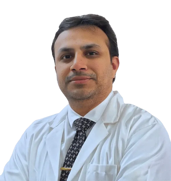
Dr. Mayur Rabhadiya
Robotic Knee & Hip Replacement Surgeon

Dr. Mandar Agashe
Paediatric Orthopedics
Our Teams at Shree Hospitals is board-certified and has an average of 15+ years of clinical experience

For Appointment/Query
1800-268-4000

Tele consultation/2nd Opinion
Schedule Consultation

News & Updates
Shree Hospitals is the first to perform robotic endo surgery.
Shree Hospitals Introduces Advanced Robotic Surgery for Better Patient Care
Shree Hospitals Expands Advanced Surgical Care With Latest Medical Technology


