 Ghatkopar (East), Mumbai)

Home > Orthopedics > Osteoporosis
Osteoporosis
At Shree Hospitals, we combine advanced hip imaging with comprehensive orthopaedic expertise to diagnose and treat hip disorders in Mumbai from early-stage conditions such as femoroacetabular impingement and hip dysplasia to advanced disease including hip osteoarthritis, avascular necrosis of the hip, and complex fractures requiring hip replacement surgery.
Our dedicated hip team orthopaedic surgeons, sports medicine physicians, and rehabilitation specialists provides an accurate diagnosis and a personalised treatment pathway focused on restoring pain-free, full hip function.
Persistent groin or deep hip pain that worsens with walking, climbing stairs, or prolonged sitting; a limp; reduced ability to rotate or flex the hip; and a grinding sensation within the joint are common signs of hip pain from structural or degenerative causes. Early specialist evaluation prevents minor hip conditions from progressing to irreversible joint damage.
(24×7 Emergency Care)
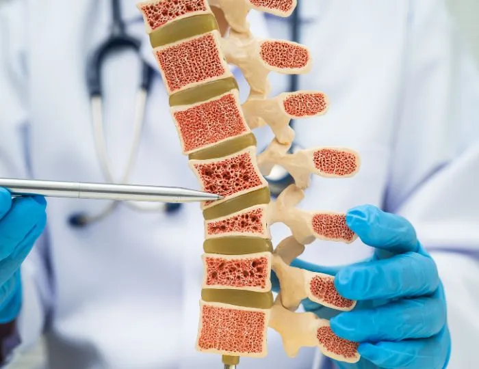
Our Approach
(24×7 Emergency Care)
Our Approach In Managing Osteoporosis
Diagnose Early. Strengthen Bone. Prevent Fractures.
At Shree Hospitals, we combine state-of-the-art bone density measurement with evidence-based medical therapy and targeted orthopaedic care to manage osteoporosis in Mumbai across all patient groups from perimenopausal women experiencing rapid early bone loss to elderly men with steroid-induced osteoporosis and to younger patients with secondary causes including malabsorption, inflammatory arthritis, and premature menopause.
Our bone health team treats osteoporosis as the serious, life-limiting medical condition it is not as a normal part of ageing to be accepted.
The consequences of untreated osteoporosis are severe. A hip fracture in a patient over 70 carries a 20–30% one-year mortality rate comparable to many cancers. Vertebral compression fractures cause chronic pain, progressive kyphotic deformity, and significantly impaired quality of life.
Yet osteoporosis remains profoundly undertreated most patients receive their first diagnosis only after sustaining a fragility fracture, by which point bone loss is already advanced. Our approach is proactive: identifying at-risk patients through DEXA scan screening, initiating appropriate bisphosphonate treatment or bone-building therapy early, and implementing comprehensive fracture prevention strategies that protect every patient from preventable injury.
- DEXA scan (dual-energy X-ray absorptiometry) at the lumbar spine and hip — the gold standard for bone mineral density (BMD) measurement and osteoporosis diagnosis
- FRAX® (Fracture Risk Assessment Tool) calculation — 10-year probability of major osteoporotic and hip fracture, integrating BMD with clinical risk factors
- Full secondary osteoporosis screen — thyroid, parathyroid, vitamin D, calcium, renal, liver, and bone turnover markers
- Vertebral fracture assessment (VFA) on DEXA — identifies prevalent vertebral compression fractures that are silent but significantly elevate future fracture risk
- Trabecular Bone Score (TBS) — assesses bone microarchitecture quality independent of BMD
- Spinal X-rays for height loss or back pain to identify morphometric vertebral fractures
Evidence from international osteoporosis guidelines confirms that patients who sustain a fragility fracture without subsequent bone health assessment and treatment have a 2–3× higher risk of a second fracture within the following 2 years (International Osteoporosis Foundation, 2024).
Our Fracture Liaison Service (FLS) systematically identifies every patient admitted with a fragility fracture and initiates secondary prevention treatment before discharge.
- Personalised treatment plans based on fracture risk category, secondary causes, patient preferences, and co-morbidities
- Choice of oral, injection-based, or intravenous bone-strengthening medications
Supervised physiotherapy and fall prevention programme - Nutritional optimisation — calcium and vitamin D therapy as the non-negotiable foundation of all treatment
- Annual DEXA monitoring to assess treatment response and adjust therapy
Our bone health clinic provides a one-stop service — DEXA scanning, specialist consultation, medication initiation, and physiotherapy referral all within a single appointment pathway.
1. Advanced Diagnosis — DEXA Scan & Bone Health Assessment
Osteoporosis diagnosis requires objective measurement of bone mineral density (BMD) clinical symptoms alone are insufficient and unreliable. Our diagnostic protocol follows the WHO and International Osteoporosis Foundation (IOF) standards.
DEXA Scan (Dual-Energy X-ray Absorptiometry):
- Measures BMD at the lumbar spine (L1–L4) and proximal femur (total hip and femoral neck) — the two most clinically relevant sites
- Results reported as T-score (comparison with young adult peak bone mass):
- T-score ≥ −1.0: Normal bone density
- T-score −1.0 to −2.5: Osteopenia (low bone density — pre-osteoporosis)
- T-score ≤ −2.5: Osteoporosis
- T-score ≤ −2.5 with one or more fragility fractures: Severe (established) osteoporosis
- Z-score (comparison with age-matched peers) — used in premenopausal women, men under 50, and children; a Z-score ≤ −2.0 indicates "below expected range for age" and mandates secondary cause investigation
- Forearm DEXA added for hyperparathyroidism, very obese patients, or where spine/hip are technically non-assessable
- Repeat DEXA every 1–2 years on treatment; every 2–3 years for monitoring in stable patients
FRAX® Fracture Risk Calculation:
- Validated WHO tool calculating 10-year probability of hip fracture and major osteoporotic fracture (spine, hip, wrist, humerus)
- Integrates BMD T-score with 11 clinical risk factors: age, sex, BMI, prior fragility fracture, parental hip fracture, current smoking, glucocorticoid use, rheumatoid arthritis, secondary osteoporosis, alcohol ≥3 units/day
- Treatment thresholds: high fracture risk (FRAX hip ≥3% or major fracture ≥20%) — initiate pharmacological therapy
- Identifies patients at high fracture risk despite BMD in the osteopenic range — avoiding under-treatment
Secondary Osteoporosis Screen (Blood Tests):
- Serum calcium, phosphate, and alkaline phosphatase (ALP)
- 25-hydroxyvitamin D — deficiency is near-universal in osteoporosis patients and must be corrected before or alongside anti-resorptive therapy
- Parathyroid hormone (PTH) — elevated in primary hyperparathyroidism, a major secondary cause
- Thyroid function tests (TSH) — hyperthyroidism accelerates bone turnover
- Full blood count and ESR — anaemia and elevated ESR suggest myeloma (a malignancy mimicking osteoporosis)
- Serum protein electrophoresis and urine Bence Jones protein — to exclude myeloma in at-risk patients
- Renal and liver function — both affect bone metabolism and drug choice
- Testosterone (men), oestradiol, FSH/LH (premenopausal women with amenorrhoea)
- Coeliac serology (anti-tTG IgA) — malabsorption is an under-recognised secondary cause
Bone Turnover Markers:
- Serum CTX (C-terminal telopeptide) — marker of bone resorption; elevated in untreated osteoporosis, myeloma, and Paget's disease; suppressed by anti-resorptive therapy (confirms treatment efficacy)
- Serum P1NP (procollagen type 1 N-terminal propeptide) — marker of bone formation; elevated by anabolic therapy (teriparatide, romosozumab); used to monitor response
2. Nutritional & Lifestyle Management
Calcium, vitamin D, and bone-loading exercise are the non-negotiable foundation of osteoporosis management — without them, pharmacological therapy is significantly less effective. No drug works optimally in a calcium- or vitamin D-deficient patient.
Calcium:
- Recommended daily intake: 1,000 mg/day (adults under 50) and 1,200 mg/day (women over 50 and all adults over 70)
- Dietary sources prioritised over supplements — dairy products, fortified plant milks, green leafy vegetables (kale, broccoli), tofu, and sesame seeds
- Calcium carbonate supplementation (taken with food) or calcium citrate (can be taken fasting, preferred post-gastric surgery) for patients who cannot meet requirements through diet
- Calcium intake above 1,500 mg/day from supplements — no additional bone benefit; possible cardiovascular concerns with excessive supplementation
Vitamin D:
- Target serum 25-hydroxyvitamin D: ≥75 nmol/L (30 ng/mL) for optimal bone health; ≥50 nmol/L as minimum
- Daily supplementation: 800–2,000 IU cholecalciferol (D3) for most adults; higher doses (3,000–5,000 IU) for deficient patients until repletion
- Monthly or quarterly high-dose loading in severely deficient patients (25,000–300,000 IU oral or IM)
- Vitamin D toxicity (>250 nmol/L serum level) is rare but monitored annually in patients on high-dose supplementation
Exercise:
- Weight-bearing and resistance exercise are the only forms of exercise proven to maintain and modestly improve BMD — swimming and cycling do not load bone sufficiently to stimulate remodelling
- Recommended programme: 30 minutes of weight-bearing activity (walking, dancing, stair climbing) 5 days per week, combined with resistance training (weights, resistance bands) 2–3 times per week
- Progressive resistance training of the hip extensors and back extensors specifically proven to reduce vertebral and hip fracture risk
- Balance training (Tai Chi, single-leg standing, perturbation training) — addresses the fall risk component of fracture prevention
- High-impact exercise (jumping, jogging) in pre-osteoporosis patients with good bone quality; avoided in established osteoporosis with vertebral fractures
Lifestyle Modification:
- Smoking cessation — smoking reduces oestrogen levels, impairs calcium absorption, and directly inhibits osteoblast function; fracture risk in smokers is 25% higher than in non-smokers
- Alcohol reduction to ≤2 units/day — alcohol inhibits osteoblasts, reduces calcium absorption, impairs vitamin D activation, and elevates fall risk
- Review of bone-harmful medications — glucocorticoids (the single biggest preventable cause of secondary osteoporosis), proton pump inhibitors, aromatase inhibitors, anticonvulsants, thiazolidinediones, and GnRH agonists
- Optimisation of underlying conditions driving bone loss — coeliac disease, inflammatory bowel disease, rheumatoid arthritis, hypogonadism
3. Medical Therapy — Bone-Strengthening Medications
Pharmacological therapy is indicated for patients with osteoporosis (T-score ≤ −2.5), high FRAX fracture risk, or a prior fragility fracture. The choice of agent depends on fracture risk level, prior fracture sites, patient preference, renal function, and co-morbidities.
Anti-Resorptive Therapy — First Line:
Bisphosphonates:
- The most widely prescribed class of osteoporosis medication worldwide — inhibit osteoclast-mediated bone resorption, preserving bone microarchitecture and reducing fracture risk
- Alendronate (Fosamax) — 70 mg once weekly oral; reduces vertebral fracture risk by 47%, hip fracture risk by 51% (FIT trial). First-line choice for most patients. Must be taken fasting with plain water and patient remains upright for 30 minutes — to prevent oesophageal irritation
- Risedronate (Actonel) — 35 mg once weekly or 150 mg once monthly; similar efficacy to alendronate; better GI tolerability
- Ibandronate (Boniva) — 150 mg once monthly oral or 3 mg IV every 3 months; reduces vertebral but not non-vertebral fractures; option when weekly tablets are not tolerated
- Zoledronic Acid (Aclasta / Reclast) — 5 mg IV infusion once yearly; the most potent bisphosphonate; reduces vertebral fracture by 70%, hip fracture by 41% (HORIZON trial); ideal for patients with poor oral medication compliance, GI disease, or following hip fracture hospitalisation
- Bisphosphonate drug holidays (after 3–5 years): residual drug in bone provides ongoing protection; holiday reduces the rare risk of atypical femoral fracture and osteonecrosis of the jaw
Denosumab (Prolia):
- A monoclonal antibody against RANKL — inhibits osteoclast formation and activity; 60 mg subcutaneous injection every 6 months
- Reduces vertebral fracture by 68%, hip fracture by 40%, non-vertebral fracture by 20% (FREEDOM trial)
- No renal dose adjustment required — preferred over bisphosphonates in moderate-severe renal impairment (eGFR <30)
- Important: must not be stopped abruptly — rapid bone loss and rebound vertebral fractures occur if doses are missed without transitioning to a bisphosphonate
- Ideal for patients who cannot take or tolerate oral bisphosphonates, or who need a twice-yearly injectable option
Anabolic (Bone-Building) Therapy — High-Risk Patients:
Teriparatide (Forteo):
- Recombinant PTH(1-34) — stimulates osteoblast activity and new bone formation (anabolic mechanism), unlike anti-resorptive agents which prevent bone loss
- 20 mcg subcutaneous daily injection for 24 months
- Reduces vertebral fracture risk by 65%, non-vertebral by 53% — superior efficacy in severe osteoporosis with multiple prior vertebral fractures
- Reserved for patients at very high fracture risk, those who have failed anti-resorptive therapy, or those with glucocorticoid-induced osteoporosis
- Must be followed by an anti-resorptive agent after the 24-month course to preserve the gains
Abaloparatide (Tymlos):
- PTHrP analogue with similar anabolic mechanism to teriparatide; 80 mcg daily subcutaneous injection for 18 months
- Slightly faster onset of BMD gain; reduced hypercalcaemia risk compared to teriparatide
Romosozumab (Evenity):
- Sclerostin inhibitor — unique dual mechanism: stimulates bone formation AND inhibits bone resorption simultaneously
- 210 mg subcutaneous injection monthly for 12 months
- Most rapid and substantial BMD gains of any osteoporosis agent — 73% reduction in new vertebral fractures vs. placebo (FRAME trial); superior to alendronate in fracture prevention (ARCH trial)
- Reserved for patients at very high fracture risk — particularly those with prior hip fracture or multiple vertebral fractures
- Contraindicated within 12 months of myocardial infarction or stroke — due to a small cardiovascular signal in the ARCH trial
- Must be followed by anti-resorptive therapy after the 12-month course
Hormone Replacement Therapy (HRT):
- Oestrogen-based HRT is highly effective at preventing bone loss in perimenopausal and early postmenopausal women — particularly where menopausal symptoms co-exist with bone loss
- Not a first-line osteoporosis treatment in older women — cardiovascular, thromboembolic, and breast cancer risk considerations limit long-term use
- Testosterone replacement in hypogonadal men — significantly improves BMD; testosterone gel or injection under endocrinology co-management
4. Fracture Management & Vertebral Augmentation
When osteoporosis-related fractures occur particularly vertebral compression fractures and hip fractures prompt specialist intervention minimises pain, prevents deformity, and enables the fastest possible recovery.
Vertebral Compression Fracture Management:
- The most common osteoporotic fracture — approximately 1.4 million new vertebral fractures occur worldwide annually
- Most are managed conservatively: analgesia, short-term rest, thoracolumbar spinal orthosis (TLSO brace), and early mobilisation
- Persistent severe pain (>4–6 weeks) unresponsive to conservative management is the primary indication for vertebral augmentation
Vertebroplasty:
- Percutaneous injection of polymethylmethacrylate (PMMA) bone cement directly into the fractured vertebral body under fluoroscopic or CT guidance
- Stabilises the fracture, eliminates pain from micromotion, and provides immediate structural support
- Performed under local anaesthesia with sedation — day-care procedure; patient mobilised within hours
- Rapid and dramatic pain relief in 80–90% of patients — typically within 24–48 hours
Balloon Kyphoplasty:
- A refinement of vertebroplasty — an inflatable balloon tamp is inserted into the fractured vertebra and inflated to restore vertebral body height before cement injection
- Restores partial vertebral height, reduces kyphotic angulation, and creates a lower-pressure cavity for cement injection — reducing cement leakage risk compared to vertebroplasty
- Preferred for sub-acute fractures (2–6 weeks old) with height loss and kyphotic deformity
- Day-care or overnight stay procedure under local anaesthesia with sedation or general anaesthesia
Hip Fracture Surgery:
- Femoral neck fractures and intertrochanteric fractures in osteoporotic patients require urgent surgical fixation (within 36–48 hours of admission) — delay is directly associated with increased mortality
- Internal fixation (dynamic hip screw, cephalomedullary nail) for stable, reducible fractures
- Hemiarthroplasty or total hip replacement for displaced femoral neck fractures — particularly in patients over 65 where bone quality prevents reliable internal fixation
- Enhanced recovery protocols and early mobilisation (Day 1 post-surgery) to minimise immobility-related complications
- Every hip fracture patient is assessed by our Fracture Liaison Service before discharge — bone health treatment initiated in hospital
5. Long-Term Monitoring & Fall Prevention
Fracture prevention in osteoporosis is a two-part strategy: strengthening bone AND preventing falls. Bone strength alone is insufficient if patients continue to fall most hip fractures result from falls, not from the bone being too weak to withstand normal loading.
Medication Monitoring:
- Annual DEXA scan for the first 2–3 years of treatment — to confirm BMD is stable or improving
- Bone turnover markers at 3 and 12 months — CTX suppression confirms anti-resorptive therapy is working; P1NP rise confirms anabolic therapy is working
- Annual blood tests: serum calcium (hypercalcaemia on teriparatide), vitamin D level, renal function (bisphosphonates), and secondary osteoporosis markers
- Bisphosphonate drug holiday assessment after 3–5 years — continuing vs. holiday decision based on fracture risk recalculation
- Transition planning: all anabolic therapy (teriparatide, romosozumab) must be followed by an anti-resorptive agent; all denosumab courses must be transitioned to bisphosphonate on cessation
Fall Prevention Programme:
- Comprehensive fall risk assessment — Timed Up and Go (TUG) test, Berg Balance Scale, orthostatic blood pressure measurement
- Balance and strengthening physiotherapy — Tai Chi (level 1 evidence for fall reduction), progressive resistance training, vestibular rehabilitation
- Home hazard assessment — removing loose rugs, improving lighting, installing grab rails and non-slip mats, raising toilet seat and chair heights
- Footwear assessment — appropriate supportive shoes; referral for hip protector devices in very high-fall-risk elderly patients
- Medication review — identifying and rationalising bone-harmful and fall-risk-increasing medications (benzodiazepines, sedatives, antihypertensives causing postural hypotension, diuretics)
- Vision correction — cataract surgery referral where poor vision is contributing to fall risk
- Cardiac assessment — syncope and postural hypotension as reversible fall causes
Patient Education:
- Understanding osteoporosis as a chronic, manageable condition — not an inevitable consequence of ageing
- Correct medication administration technique (particularly for weekly bisphosphonates and self-injection with teriparatide or denosumab)
- Recognising vertebral fracture symptoms — acute back pain after minimal exertion, height loss measurement at home
- When to seek emergency care — sudden severe back pain, inability to weight-bear after a fall, neurological symptoms after a vertebral fracture
Happy Patients & Their Case Stories
From diagnosis to discharge a 62-year-old postmenopausal woman presented to Shree Hospitals after sustaining a wrist fracture following a minor fall. DEXA scan confirmed severe osteoporosis (T-score −3.1 at the lumbar spine). She was commenced on zoledronic acid infusion, calcium, and vitamin D supplementation alongside a supervised balance and strengthening programme. At 2-year follow-up, her lumbar spine BMD had improved by 8% and she has had no further fractures.
From diagnosis to discharge a 74-year-old man with chronic back pain and 4 cm height loss was found to have three silent vertebral compression fractures on vertebral fracture assessment. He underwent bilateral balloon kyphoplasty at T12 and L1, achieving complete pain relief within 48 hours. He was commenced on teriparatide followed by denosumab for ongoing bone protection.
How to identify Osteoporosis?
Osteoporosis is called the "silent disease" because it causes no symptoms until a fracture occurs by which point significant bone loss has already taken place. The fracture itself is often the first and most devastating sign.
The most important warning sign of osteoporosis is a fracture from a low-energy injury breaking a wrist from a fall from standing height, a vertebral fracture from sneezing or coughing, or a hip fracture from a simple trip.
Any fracture in an adult over 50 from minimal trauma must be investigated as a potential osteoporotic (fragility) fracture.
Key warning signs and risk indicators to be aware of:
- Fragility fracture — a fracture caused by a force that would not normally break a healthy bone; falling from standing height is the standard threshold; a prior fragility fracture doubles the risk of the next one
- Back pain following minor strain — sudden onset of mid or lower back pain after bending, lifting, or even coughing may indicate a silent vertebral compression fracture
- Height loss of 2 cm or more compared to peak adult height — each vertebral compression fracture typically causes 1–2 cm of height loss; cumulative height loss of 4 cm or more strongly suggests multiple vertebral fractures
- Progressive stoop or dowager's hump — increasing thoracic kyphosis (rounded upper back) from multiple vertebral compression fractures stacking on each other
Loss of teeth or receding gum line in older patients — may reflect systemic bone density loss, though jaw bone loss has a different aetiology - Risk factors in the absence of symptoms — postmenopausal status, age over 65 (women) or 70 (men), long-term steroid use (>3 months at ≥5 mg prednisolone/day), family history of hip fracture, low BMI (<19 kg/m²), smoking, heavy alcohol use, rheumatoid arthritis, malabsorption conditions
Screening DEXA scan is recommended for all women over 65, all men over 70, and for younger patients with any significant risk factor. Do not wait for a fracture to investigate bone density.
Important FAQs: Osteoporosis
What is osteoporosis and how does it differ from osteopenia?
Osteoporosis is a systemic skeletal disease characterised by reduced bone mass and deterioration of bone microarchitecture, resulting in increased bone fragility and fracture risk. It is diagnosed when the bone mineral density T-score is −2.5 or below on DEXA scan. Osteopenia is an intermediate state T-score between −1.0 and −2.5 where bone density is below normal but has not yet reached the osteoporosis threshold. Osteopenia is not a disease but a risk category many patients with osteopenia have a high enough FRAX fracture risk to warrant pharmacological treatment, while others can be managed with lifestyle measures alone. Both conditions are part of the same spectrum of bone density loss and require assessment rather than dismissal.
Who is most at risk of developing osteoporosis?
The highest-risk groups are: postmenopausal women (oestrogen withdrawal accelerates bone loss by 2–3% per year in the first 5–10 years after menopause), adults over 65 (men and women), patients taking long-term oral corticosteroids (≥5 mg prednisolone daily for ≥3 months — the most common cause of secondary osteoporosis, and one of the most undertreated), patients with rheumatoid arthritis, inflammatory bowel disease, coeliac disease, or other malabsorption conditions, individuals with a family history of hip fracture, smokers, heavy alcohol users, those with low BMI, and anyone who has already sustained a fragility fracture. In India specifically, vitamin D deficiency is near-universal and contributes significantly to poor bone health across all age groups.
How is osteoporosis diagnosed at the hospital?
Diagnosis requires a DEXA scan — measuring bone mineral density at the lumbar spine and hip — to determine the T-score. FRAX® fracture risk calculation integrates BMD with clinical risk factors to produce a 10-year fracture probability, which directly informs treatment decisions. A full secondary osteoporosis blood screen (vitamin D, calcium, PTH, thyroid, renal and liver function, blood count, myeloma screen) is performed to identify and treat correctable underlying causes. Vertebral fracture assessment on the DEXA identifies silent vertebral fractures that elevate fracture risk independent of BMD.
Can osteoporosis be reversed with treatment?
Osteoporosis cannot be fully "cured" bone lost through years of progressive thinning cannot be completely restored. However, all currently available treatments can significantly slow or halt further bone loss, and some agents (teriparatide, romosozumab) can rebuild meaningful amounts of new bone, improving T-score by 0.5–1.0 SD or more. More importantly, all proven treatments substantially reduce fracture risk by 40–70% depending on the agent which is the primary clinical goal. With appropriate treatment, monitoring, and lifestyle modification, most patients with osteoporosis can live fracture-free, active, independent lives for decades.
What medications are used for osteoporosis and how long must they be taken?
First-line treatment is a bisphosphonate (alendronate, risedronate, or zoledronic acid) — taken for 3–5 years, after which a drug holiday is considered based on ongoing fracture risk assessment. Denosumab is given indefinitely (as long as treatment is indicated) but must be transitioned carefully to a bisphosphonate if stopped. Teriparatide and romosozumab are anabolic agents used for 12–24 months in high-risk patients, always followed by anti-resorptive therapy to preserve gains. Calcium and vitamin D are lifelong supplements for all patients. Osteoporosis is a chronic condition — treatment should be viewed as long-term management, not a short course.
What is balloon kyphoplasty and who needs it?
Balloon kyphoplasty is a minimally invasive procedure for painful vertebral compression fractures — the most common fracture type in osteoporosis, affecting approximately 700,000 patients in India annually. Under local anaesthesia and sedation, a small balloon is inserted into the collapsed vertebra through a needle and inflated to partially restore vertebral height, then deflated and the cavity filled with bone cement to stabilise the fracture permanently. The procedure takes 30–60 minutes per vertebra and is performed as a day-care or overnight stay. It provides rapid, often immediate, pain relief in 80–90% of patients and is indicated when back pain from a vertebral fracture is severe and has not responded to 4–6 weeks of conservative management.
When should I get a DEXA scan or see a bone health specialist?
A DEXA scan is recommended for all women over 65 and men over 70 regardless of symptoms. It is recommended earlier for: any adult who has sustained a fragility fracture (wrist, vertebra, hip, or humerus from minimal trauma), postmenopausal women under 65 with one or more risk factors, men aged 50–70 with significant risk factors, anyone on long-term steroids, patients with conditions associated with bone loss (rheumatoid arthritis, coeliac disease, inflammatory bowel disease, primary hyperparathyroidism), and those with a family history of hip fracture. Do not wait for back pain or a fracture to seek evaluation osteoporosis is eminently preventable if identified and treated before the first fracture occurs.
Treatments for Osteoporosis
at Shree Hospitals
We provide comprehensive treatment for osteoporosis, including accurate diagnosis, personalised treatment plans, advanced medical management, fracture prevention strategies, bone health optimisation, rehabilitation, and follow-up care to improve bone strength, reduce fracture risk, relieve pain, enhance mobility, and improve overall quality of life.
DEXA Scan & Bone Density Assessment
Gold-standard dual-energy X-ray absorptiometry measuring bone mineral density at the spine and hip with FRAX® fracture risk calculation, vertebral fracture assessment, and full secondary osteoporosis blood screen in a single clinic visit.
Calcium & Vitamin D Therapy
The non-negotiable foundation of all osteoporosis treatment dietary calcium optimisation, targeted supplementation to reach 1,000–1,200 mg/day, and vitamin D therapy to achieve serum levels ≥75 nmol/L for optimal bone health.
Bisphosphonate Therapy (Oral & IV)
First-line anti-resorptive medication weekly oral alendronate or risedronate, or annual zoledronic acid IV infusion proven to reduce vertebral fracture risk by up to 70% and hip fracture risk by over 40%.
Denosumab Injections
Six-monthly subcutaneous injection targeting RANKL to inhibit osteoclast bone resorption preferred for patients with renal impairment, GI intolerance to bisphosphonates, or who need an injectable option with superior BMD gains.
Teriparatide (Bone-Building Therapy)
Daily subcutaneous injection of recombinant PTH for 24 months anabolic therapy that stimulates new bone formation; reserved for severe osteoporosis, multiple vertebral fractures, and treatment-failed cases.
Romosozumab (Dual-Action Therapy)
Monthly sclerostin inhibitor injection simultaneously builds new bone and prevents resorption; the most potent single-agent BMD improvement available; used for patients at very high fracture risk.
Balloon Kyphoplasty / Vertebroplasty
Percutaneous cement injection into painful osteoporotic vertebral compression fractures restores vertebral height, eliminates fracture pain, and stabilises the spine; performed as a day-care procedure with rapid recovery.
Fall Prevention & Physiotherapy Programme
Comprehensive supervised programme combining Tai Chi-based balance training, progressive resistance exercise for hip and spine strength, home hazard assessment, and medication review addressing the fall risk side of fracture prevention.
Top Orthopaedic Specialist in Mumbai
Dr. Tapan Shah
Physiotherapist

Dr. Chirag Dalal
Orthopedic Arthroscopy & Trauma Surgeon

Dr. Mayur Rabhadiya
Robotic Knee & Hip Replacement Surgeon

Dr. Mandar Agashe
Paediatric Orthopedics
Our Teams at Shree Hospitals is board-certified and has an average of 15+ years of clinical experience

For Appointment/Query
1800-268-4000

Tele consultation/2nd Opinion
Schedule Consultation

News & Updates
Shree Hospitals is the first to perform robotic endo surgery.
Shree Hospitals Introduces Advanced Robotic Surgery for Better Patient Care
Shree Hospitals Expands Advanced Surgical Care With Latest Medical Technology
Health Center
Exploring the Future of Telemedicine: What’s Next for Healthcare?
The Rise of Renewable Energy: Innovations in Sustainability
Follow Us

Shree
Hospitals

Shree
Hospitals
