 Ghatkopar (East), Mumbai)

Home >Pulmonology > Pneumonia
Pneumonia
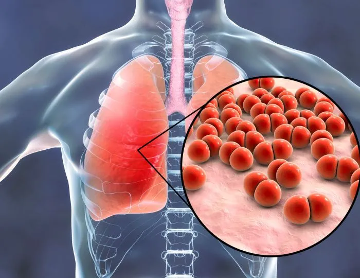
Pneumonia kills more people worldwide than any other infectious disease - and in India, it remains a leading cause of hospital admission and death across all age groups. It is not just a severe cold. Pneumonia is an infection of the lung parenchyma - the air sacs fill with fluid and pus, oxygen exchange collapses, and what looks like a manageable respiratory illness can deteriorate into sepsis and respiratory failure within 24 hours if not assessed and treated correctly. At Shree Hospitals, our pneumonia treatment in Mumbai is structured around rapid severity assessment, targeted antibiotic therapy for pneumonia, and clinical monitoring that catches deterioration before it becomes irreversible. Whether you are walking in with fever and cough or arriving in ambulance with oxygen saturation in the 80s - our respiratory team responds to the clinical picture, not a protocol alone.
(24×7 Emergency Care)
Our Approach
(24×7 Emergency Care)
Our Approach in Managing Pneumonia
The first 6 hours of pneumonia management determine the trajectory. Our pneumonia specialist pulmonologist team in Mumbai, India uses a structured triage-to-treatment protocol that does not wait for culture results before initiating appropriate empirical therapy, while simultaneously collecting the microbiological data that allows de-escalation once organisms are identified.
- Clinical Assessment & Severity Scoring
Not every pneumonia patient needs admission. Not every admitted patient is adequately monitored.
- CURB-65 score: Confusion, Urea >7 mmol/L, Respiratory rate ≥30, BP <90/60, Age ≥65 - score 0–1: outpatient; 2: consider admission; 3–5: hospital, consider ICU
- PSI (Pneumonia Severity Index) - more granular scoring for hospitalised patients guiding ICU versus ward allocation
- Vital signs: respiratory rate, SpO2, heart rate, BP, temperature - the clinical severity determinants
- Oxygen saturation trending - SpO2 below 92% on room air requires immediate escalation
- Diagnostic Workup
- Chest X-ray - confirms consolidation, identifies distribution (lobar, multilobar, interstitial), excludes complications (effusion, empyema)
- CT chest - for atypical presentations, immunocompromised patients, or suspected complications
- Blood cultures × 2 - before antibiotics in moderate to severe pneumonia
- Sputum culture and Gram stain - where patient can produce a good sample
- Pneumococcal and Legionella urinary antigen tests - rapid identification of the two most common severe CAP organisms
- Nasopharyngeal swab for respiratory viruses - influenza, COVID-19, RSV in appropriate context
- Procalcitonin and CRP - severity markers and antibiotic stewardship guides
- Antibiotic Therapy - Empirical Then Targeted
Antibiotic therapy for pneumonia must be prompt - delays beyond 4 hours worsen outcomes.
- Mild CAP (outpatient): oral Amoxicillin ± Azithromycin for atypical cover; Doxycycline as alternative
- Moderate CAP (ward): IV Beta-lactam (Ampicillin-sulbactam, Cefotaxime) + Macrolide or respiratory fluoroquinolone (Levofloxacin)
- Severe CAP (ICU): IV Beta-lactam + Macrolide or respiratory fluoroquinolone; add Vancomycin or Linezolid if MRSA risk; Piperacillin-tazobactam if Pseudomonas risk
- De-escalation at 48–72 hours based on culture results and clinical response
- Antibiotic duration: 5 days for mild-moderate CAP responding well; 7 days for severe; extended for specific organisms (Legionella, S. aureus)
- Hospital-acquired pneumonia (HAP) and ventilator-associated pneumonia (VAP): broader spectrum empirical therapy guided by local resistance patterns
- Respiratory Support
- Controlled oxygen - target SpO2 94–98% (88–92% in COPD patients)
- High-Flow Nasal Cannula (HFNC) - for hypoxaemic respiratory failure; reduces intubation rates compared to conventional oxygen
- Non-invasive ventilation (BiPAP) - for hypercapnic patients or those with underlying lung disease
- Invasive mechanical ventilation - for respiratory failure unresponsive to HFNC/NIV; lung-protective ventilation strategy (low tidal volume, PEEP)
- Prone positioning - for severe ARDS complicating pneumonia with PaO2/FiO2 <150
- Managing Complications
- Parapneumonic effusion - thoracocentesis for diagnostic and therapeutic drainage; pleural fluid pH, glucose, LDH to distinguish exudate from empyema
- Empyema - chest tube drainage; surgical decortication if organised
- Lung abscess - prolonged antibiotic therapy; bronchoscopic or percutaneous drainage for non-responding abscesses
- Septic shock - IV fluid resuscitation, vasopressors, source control, and ICU transfer
- Respiratory failure - escalation through oxygen → HFNC → NIV → intubation pathway based on clinical trajectory
- Pneumonia Recovery & Discharge Planning
Pneumonia recovery and complications monitoring extends beyond hospital discharge.
- Clinical response expected within 48–72 hours of appropriate antibiotics - failure to improve triggers reassessment
- Radiological resolution lags clinical recovery by 4–6 weeks - repeat X-ray not needed before 4 weeks in uncomplicated cases
- Post-discharge follow-up at 4–6 weeks with repeat chest X-ray - persistent opacity requires further investigation to exclude malignancy
- Pneumococcal and influenza vaccination before discharge for high-risk patients
- Rehabilitation for post-severe-pneumonia patients - fatigue, reduced exercise capacity, and psychological impact are common.
Happy Patients & Their Case Stories
Presented with three days of fever, productive cough, and mild confusion. SpO2 was 84% on room air. CURB-65 score was 4. CT chest showed right lower lobe consolidation with early parapneumonic effusion. He was started on IV Cefoperazone-sulbactam and Azithromycin, placed on HFNC, and transferred to our respiratory HDU.
Mr. Ashish .P
A diabetic who came to our emergency department with rapidly worsening breathlessness over 12 hours. CT chest showed bilateral multilobar consolidation. COVID-19 PCR was positive. She was started on Dexamethasone, anticoagulation, and HFNC. She did not require intubation.
Mr. Randive. L
How to Identify Pneumonia?
Pneumonia is often confused with a severe cold or a viral upper respiratory infection - the distinction matters because the management is entirely different.
Features that indicate pneumonia rather than a simple respiratory infection:
- Fever above 38.5°C with shaking chills - not typical of a cold
- Productive cough with green, yellow, or rust-coloured sputum
- Breathlessness on mild exertion or at rest - not present in an uncomplicated cold
- Pleuritic chest pain - sharp, one-sided pain that worsens on deep breathing or coughing
- SpO2 below 94% on pulse oximetry - a simple home measurement that should trigger immediate medical review
- Symptoms that are worsening after 48–72 hours rather than improving
- Confusion, drowsiness, or inability to complete sentences - indicating severe lung infection
The populations at highest risk - elderly patients, diabetics, immunocompromised patients, those with pre-existing lung disease - should have a lower threshold for seeking evaluation because pneumonia in these groups progresses faster and with less warning. Antibiotic therapy for pneumonia initiated within 4 hours of appropriate diagnosis is a documented outcome determinant. Seek pneumonia treatment in Mumbai at a centre equipped to assess severity, escalate oxygen support, and move to ICU-level care if the trajectory demands it.
Important FAQs : Pneumonia
How long does pneumonia take to recover from?
Most patients feel significantly better within 3–5 days of antibiotics. Full clinical recovery takes 1–3 weeks. Radiological resolution on chest X-ray takes 4–8 weeks and should not be used as the sole marker of recovery.
Is pneumonia contagious?
Bacterial pneumonia - the most common type - has limited person-to-person spread. Viral pneumonias, including influenza and COVID-19 pneumonia, are more readily transmissible through respiratory droplets.
When should a child with pneumonia be hospitalised?
Children with SpO2 below 92%, respiratory rate above age-normal limits, inability to feed, signs of severe respiratory distress, or failing outpatient antibiotic therapy require admission.
Can pneumonia be prevented?
Pneumococcal vaccination (PCV13, PPSV23) reduces the incidence of Streptococcus pneumoniae pneumonia significantly. Influenza vaccination reduces influenza-associated pneumonia. Smoking cessation reduces pneumonia risk substantially.
What is walking pneumonia?
Walking pneumonia - typically caused by Mycoplasma pneumoniae or Chlamydophila pneumoniae - produces milder symptoms that do not prevent the patient from being ambulatory. It requires macrolide or doxycycline therapy rather than standard beta-lactams.
Treatments for Pneumonia at Shree Hospitals
Comprehensive community acquired pneumonia management in Mumbai from outpatient mild infections to ICU-level severe pneumonia.
High-Flow Nasal Cannula (HFNC)
Advanced oxygen delivery for hypoxaemic lung infection presentations - providing heated humidified oxygen at flows up to 60L/min, reducing work of breathing and intubation rates in moderate-severe pneumonia.
Top Pulmonology & Infectious Disease Specialists in Mumbai

Dr. Jay Bhanushali
Pulmonologist

Dr. Tejal Shah
Interventional Pulmonology, Tuberculosis Management

Dr. Miti A Shah
Interventional Pulmonology, Pediatric Respiratory

Dr. Rishabh Raj
Interventional Pulmonology, Sleep Medicine
Every doctor and specialist at Shree Hospitals is board-certified and brings an average of 15 or more years of clinical experience in their area of subspecialty.

For Appointment/Query
1800-268-4000

Tele consultation/2nd Opinion
Schedule Consultation

News & Updates
Shree Hospitals is the first to perform robotic endo surgery.
Shree Hospitals Introduces Advanced Robotic Surgery for Better Patient Care
Shree Hospitals Expands Advanced Surgical Care With Latest Medical Technology


